Images
Vidéos
13674341 - RSV virus particles in human lung cells, SEM
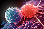
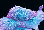
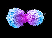
13756611 - Lung cancer cells, SEM
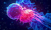
12969742 - Pancreatic cancer cell, SEM

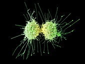
12656691 - Cervical cancer cells dividing, SEM

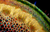
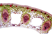
12638779 - Root tip meristem cell, TEM
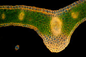
13416884 - Astrocyte cells, fluorescence micrograph
13687264 - Lung cancer cells, SEM
13471507 - Lung cancer cell, SEM
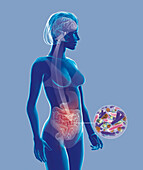
13223508 - Intestinal villi, illustration

12961105 - Organ of corti,SEM

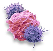
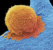
14078713 - (CAR) T-cell therapy, SEM
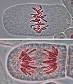
13755894 - Mitosis and cytokinesis, light micrograph
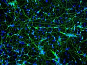
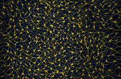
13387800 - Cortical neurons, fluorescence light micrograph

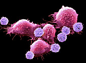
13386717 - T cells attacking cancer cells, illustration

13686807 - CAR T cell immunotherapy, illustration
12504096 - CAR T-cell immunotherapy, SEM
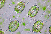
12450494 - Lily (Lilium sp.) stomata, light micrograph
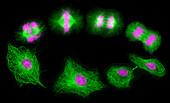
13755893 - Mitosis and cytokinesis, light micrograph
13387798 - Neural stem cells, fluorescence light micrograph
13732931 - Lymphoma, SEM

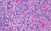
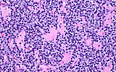
13523549 - Multiple myeloma, light micrograph
14077709 - Lung cancer cells dividing, SEM
14077649 - Dividing HeLa cells, SEM
13954484 - Nerve ending, TEM
13954451 - Nerve ending, TEM

13950881 - Brain research, conceptual illustration
13950521 - Smooth muscle cells, light micrograph
13950512 - Saponification fat necrosis, light micrograph
13950502 - C4d kidney biopsy, light micrograph
13950476 - Liver cirrhosis, light micrograph
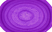
13950436 - Corpus amylaceous, light micrograph
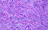
13950424 - Pleomorphic adenoma, light micrograph

13950383 - Lung cancer cells, SEM
13765641 - T cells attacking cancer cell, illustration
13756605 - Cervical cancer cells dividing, SEM
13755908 - Oat stomata, light micrograph
13755852 - Marsh-marigold stalk, light micrograph
13754734 - Mitosis in binucleate cells, light micrograph
13742779 - Lung cancer cells dividing, SEM
13732937 - Fungal infection, SEM
13732514 - Collagen substance, light micrograph
13732471 - Pacinian corpuscle, light micrograph

13732454 - Oesophagus squamous cells, light micrograph
13732425 - Normal nerve, light micrograph
13732411 - Liver ischemia, light micrograph
13732388 - Diabetic kidney disease, light micrograph
13686451 - CAR T-cell therapy, SEM

13674183 - Green alga, light micrograph
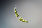
13674086 - Closterium sp. desmid, light micrograph
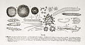
13634096 - Cells under microscope, 19th century illustration
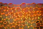
13633753 - Collenchyma in garden rhubarb stalk, light micrograph
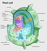
13618819 - Plant cell, illustration
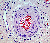
14084159 - Arteriole, LM
14078709 - (CAR) T-cell therapy, SEM
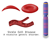
14078388 - Normal blood cell and sickle cell disease, illustration
13954159 - Stomata in hyacinth (Hyacinthus sp.) leaf, light micrograph
13951778 - Cell membrane semi-permeability, illustration
13950519 - Skin epidermis, light micrograph
13950503 - C4d kidney biopsy, light micrograph
13950477 - Liver cirrhosis, light micrograph
13950469 - Intima and media of kidney blood vessel, light micrograph
13950463 - Papillary bladder cancer, light micrograph
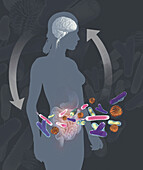
13839366 - Gut brain connection, illustration
13839362 - Gut brain connection, illustration
13838429 - Knautia sp. stalk, light micrograph
13765644 - T cell attacking cancer cell, illustration
13765642 - T cell attacking cancer cell, illustration
13756616 - Prostate cancer cells, SEM
13756606 - Cervical cancer cells dividing, SEM
13755863 - Lily of the valley leaf, light micrograph
13755855 - Marsh-marigold leaf, light micrograph
13754701 - Mitosis, light micrograph
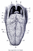
13736195 - Tongue anatomy, illustration
13732510 - Nerve ganglion cells, light micrograph
13732504 - Substance in Warthin tumour, light micrograph
13732398 - Uterus endometrium with atrophy, light micrograph
13732355 - Smooth muscle cells in stomach, light micrograph

13732349 - Fat cells, light micrograph
13686210 - Compact bone, light micrograph
13686100 - Newt larvae ciliated cells, SEM
13634175 - Testis, SEM
13634090 - Cells under microscope, 19th century illustration

13633179 - Red blood cells, SEM
13633142 - Pluripotent derived neurones, SEM
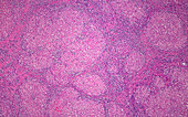
13601249 - Cirrhosis, light micrograph
13525553 - Yellow fever virus, TEM

13377010 - CAR T cell immunotherapy, illustration
13243219 - Single cortical neuron, light micrograph
12971158 - Prostate cancer cells, SEM
14084180 - Ulcerative colitis, LM

14084175 - Inflammation, LM
14084174 - Cervical cancer, LM
14084173 - Inflammation, LM

14078405 - Sickle cell disease inheritance, illustration
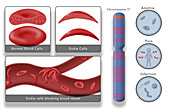
14078383 - Sickle cell disease, illustration
14077661 - Dividing HeLa cells, SEM
13952289 - Prostate cancer cells dividing, SEM
13950835 - Neurons, conceptual illustration
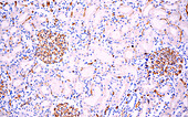
13950495 - Parathyroid gland cells, light micrograph
page suivante
Cellules Photos ❘ Science Photo Library