Iris layers, light micrograph
Numéro d’image : 12949078
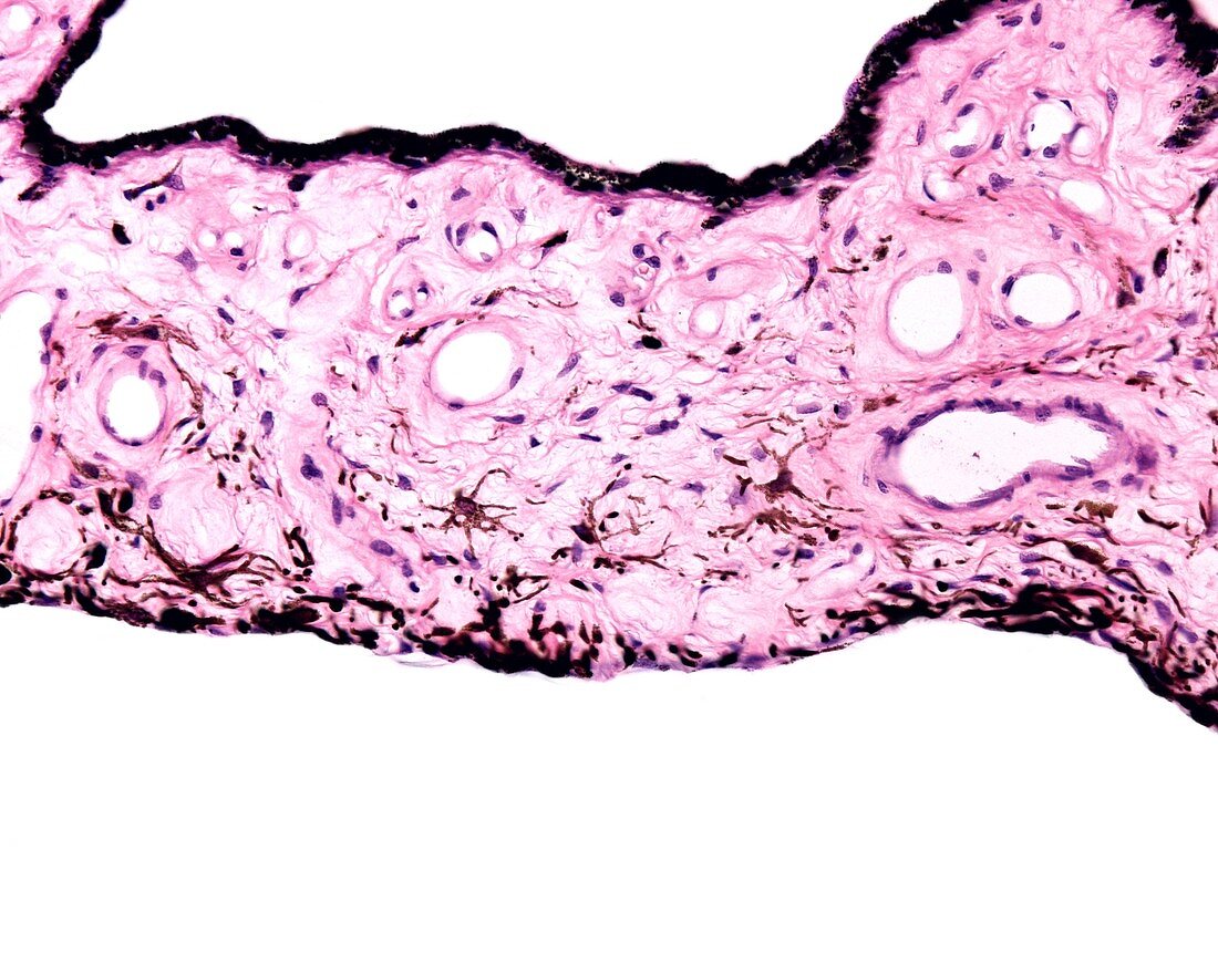
| Iris layers, light micrograph. From bottom to top: anterior chamber, anterior surface (without epithelium), iris stroma with many pigment cells and blood vessels, anterior and posterior pigmented epitheliums and posterior chamber. | |
| Licence : | Droits gérés |
| Crédit: | Science Photo Library / JOSE CALVO |
| Taille de l’image : | 3840 px × 3072 px |
| Model Release : | Non requis |
| Property Release : | Non requis |
| Restrictions : | - |
Prix pour cette image À partir de 45 €
Produit vendu
(Calendrier, Carte postale, Carte de vœux, Impression sur textile, Packaging etc)
À partir de 45 €
Usage commercial
(Affichage, Annonce presse, Annonce TV, Carte, Digital - hors rés. sociaux, Digital - rés. sociaux etc)
À partir de 45 €
Éditorial
(Digital, Journal, Livre, Livre pratique, Magazine, Télévision etc)
À partir de 60 €
Usage non-commercial
(Digital - hors rés. sociaux, Digital - rés. sociaux etc)
À partir de 120 €
Mots clés
- aucun,
- biologie,
- biologique,
- coloré,
- epithelium,
- épithélium,
- globe oculaire,
- histologie,
- histologique,
- iris,
- mélanocytes,
- micrographie,
- micrographie optique,
- microscope,
- microscope optique,
- microscopie,
- microscopie optique,
- microscopique,
- oculaire,
- oeil,
- ophtalmique,
- ophtalmologie,
- personne,
- pigmenté