Images
Vidéos
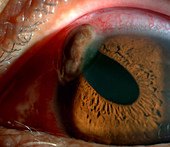
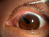
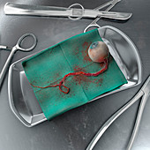
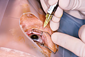
12629332 - Ruptured Globe
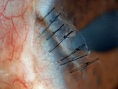
12629334 - Repaired Ruptured Globe
12629331 - Ruptured Globe
12037009 - Punctured Globe,Ophthalmic Medicine
12036938 - Ruptured Globe
12036939 - Ruptured Globe
12037010 - Punctured Globe,Ophthalmic Medicine
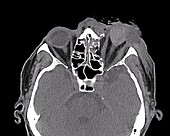
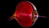
13674983 - Ruptured globe, CT scan
12629333 - Repaired Ruptured Globe
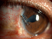
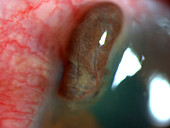
12036671 - Punctured Globe
12036674 - Punctured Globe
12036673 - Punctured Globe
12036672 - Punctured Globe
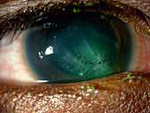
12036931 - Enucleated Eye
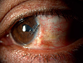
11844662 - Eye injury
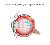
13736725 - Retinal detachment, illustration
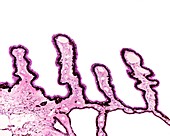
13756214 - Schlemm canal in eye, light micrograph

13736724 - Retinal detachment, illustration
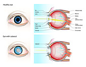
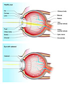
13736718 - Normal eye and eye with cataract, illustration

13736716 - Healthy eye and diabetic retinopathy, illustration
13756213 - Choroid and retina, light micrograph

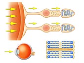
13585907 - Light entering human eye, illustration
13756200 - Eye lens fibres, light micrograph
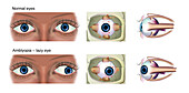
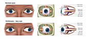
13736714 - Normal eye and amblyopia, illustration
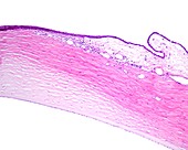
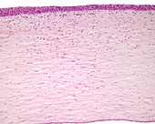
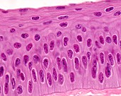
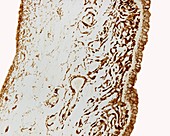
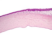
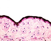
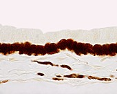
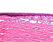
13756183 - Human cornea, light micrograph
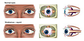
13736727 - Normal eye and strabismus, illustration

13736715 - Healthy eye and diabetic retinopathy, illustration
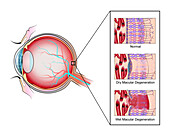
13736723 - Macular degeneration, illustration

13736722 - Macular degeneration, illustration
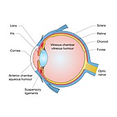
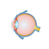
13585904 - Human eye anatomy, illustration
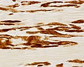
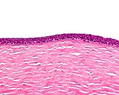
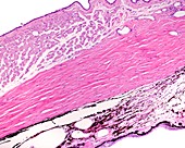
13426612 - Conjunctiva and sclera, light micrograph
13377636 - Eye anatomy, illustration

13756179 - Human cornea, light micrograph
13416647 - Iris, light micrograph
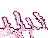
13756215 - Human ciliary body and iris, light micrograph
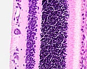
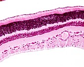
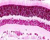
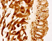
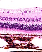
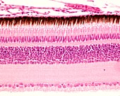
13426632 - Retina layers, light micrograph
13243917 - Ciliary body epithelium, light micrograph
13243908 - Iris stroma, light micrograph

13426627 - Iris sphincter muscle, light micrograph
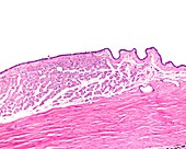
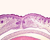
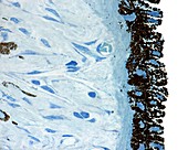
13426623 - Limit between cornea and conjunctiva, light micrograph
13243915 - Retina layers, light micrograph
13736713 - Normal eye and amblyopia, illustration
13426624 - Ciliary body, light micrograph

13376764 - Eye anatomy, illustration
13243896 - Cornea layers, light micrograph
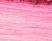
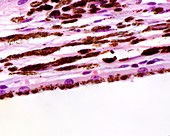
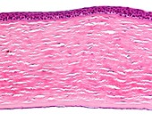
13243889 - Sclera, light micrograph
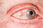
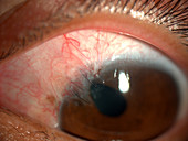
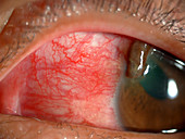
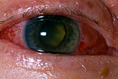
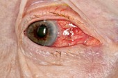
13951631 - Inflamed white of the eye
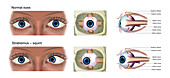
13736726 - Normal eye and strabismus, illustration
13736721 - Normal eye and eye with cataract, illustration
13243905 - Cornea epithelium, light micrograph
13243900 - Choroid layer, light micrograph
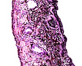
13243886 - Eye development, light micrograph
12987500 - Eye lens, light micrograph
13736719 - Normal eye and eye with cataract, illustration
13756216 - Human conjunctiva and cornea, light micrograph

13736717 - Normal eye and eye with glaucoma, illustration
13243903 - Sclera, light micrograph
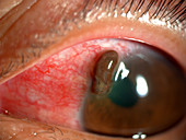
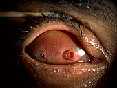
12378553 - Acute glaucoma in a blind eye

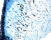
13243916 - Cat tapetum lucidum, light micrograph
13243901 - Sclera, light micrograph
13377637 - Eye anatomy, illustration
13243919 - Retina layers, light micrograph

13243897 - Cornea, light micrograph
12949097 - Cornea, light micrograph
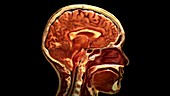
12647808 - Sagittal Section of Head
13377002 - Rear surface of iris, light micrograph

13376761 - Muscles of the eye, illustration
13243909 - Iris, light micrograph
13243907 - Back of iris, light micrograph
13243898 - Cornea and conjunctiva, light micrograph
12971338 - Biochemistry of the retina, illustration
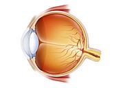
12971292 - Eye anatomy, illustration
13243914 - Lamina fusca, light micrograph
12949068 - Ciliary body, light micrograph
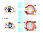
13736720 - Normal eye and eye with cataract, illustration
12949110 - Retina and choroid, light micrograph
13426625 - Ciliary processes, light micrograph
12949083 - Iris, light micrograph
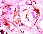
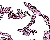
12949072 - Melanocytes in iris stroma, light micrograph
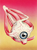
13376759 - Muscles of the eye, illustration
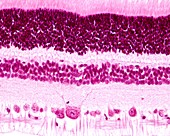
13243918 - Retina layers, light micrograph
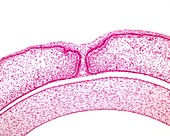
12949098 - Embryonic fused eyelids, light micrograph
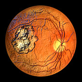
13956085 - Retinal scar caused by toxoplasmosis, illustration
13956074 - Retinal scar caused by toxoplasmosis, illustration
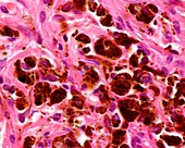
12645843 - Eye pathology examination, illustration
13243910 - Back of iris, light micrograph
13243906 - Ciliary body epithelium, light micrograph
12949077 - Iris pigment cells, light micrograph
11725642 - Macular degeneration treatment
13377003 - Rear surface of iris, light micrograph

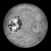
13956070 - Retinal scar and healthy retina, illustration
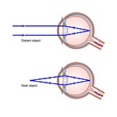
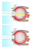
13377638 - Accommodation in the eye, illustration
13243888 - Choroid pigment cells, light micrograph
12949096 - Cornea, light micrograph
13426629 - Iris of the eye, light micrograph
13243913 - Ciliary body epithelium, light micrograph
12949109 - Layers of the retina, light micrograph
12949071 - Conjunctiva layer of eye, light micrograph
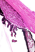
13426614 - Eye layers, light micrograph
page suivante
Globe oculaire Photos ❘ Science Photo Library