Images
Vidéos
13736725 - Retinal detachment, illustration

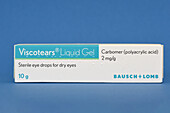
13674391 - Liquid gel eye drops
13756214 - Schlemm canal in eye, light micrograph
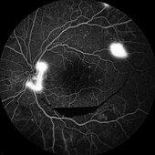
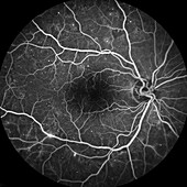
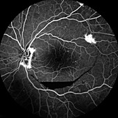
13613568 - Retina damage from diabetes, angiogram

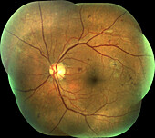
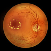
13613650 - Retina damage from diabetes, fundoscopy

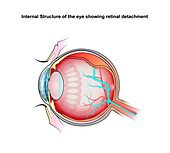
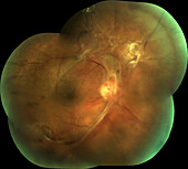
13736724 - Retinal detachment, illustration
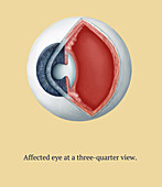
13633364 - Cat eye with hypertension, illustration
13613640 - Retina damage from diabetes, angiogram
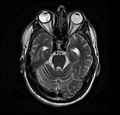
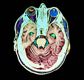
13599593 - Retinal detachment, MRI scan
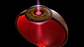
13585921 - Light entering human eye, illustration
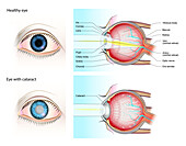
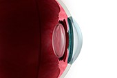
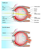
13736718 - Normal eye and eye with cataract, illustration

13736716 - Healthy eye and diabetic retinopathy, illustration

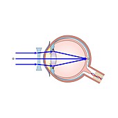
13585905 - Light entering human eye, illustration
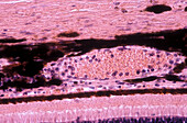
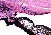
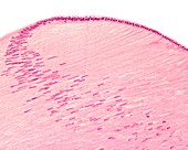
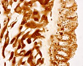
13756213 - Choroid and retina, light micrograph
13585907 - Light entering human eye, illustration
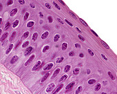
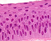
13756200 - Eye lens fibres, light micrograph
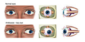
13736714 - Normal eye and amblyopia, illustration

12969886 - Kayser-Fleischer rings in eyes due to liver disease

14077544 - Glaucoma eye drops
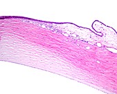
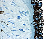
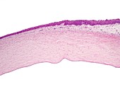
13756183 - Human cornea, light micrograph
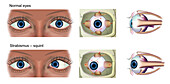
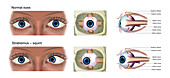
13736727 - Normal eye and strabismus, illustration

13736715 - Healthy eye and diabetic retinopathy, illustration
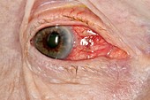
12644137 - Anterior uveitis
13613570 - Retina damage from diabetes, angiogram
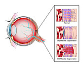
13736723 - Macular degeneration, illustration

13736722 - Macular degeneration, illustration

13613647 - Retina damage from diabetes, angiogram
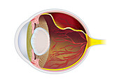
13585904 - Human eye anatomy, illustration
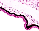
13426612 - Conjunctiva and sclera, light micrograph

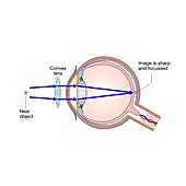
13377650 - Long-sightedness corrected, illustration
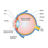
13377636 - Eye anatomy, illustration
12991282 - Eye anatomy, illustration

13756179 - Human cornea, light micrograph
13416647 - Iris, light micrograph
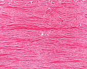
13243887 - Alveolar gland, light micrograph
13756215 - Human ciliary body and iris, light micrograph
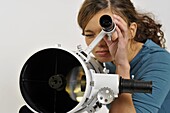
13478996 - Woman looking through eyepiece of finderscope
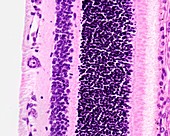
13426632 - Retina layers, light micrograph
13243917 - Ciliary body epithelium, light micrograph
13243908 - Iris stroma, light micrograph
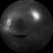
13613572 - Retina damage from diabetes, angiogram

13426627 - Iris sphincter muscle, light micrograph
13426623 - Limit between cornea and conjunctiva, light micrograph
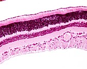
13243915 - Retina layers, light micrograph
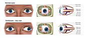
13736713 - Normal eye and amblyopia, illustration
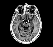
13599595 - Retinal detachment, MRI scan
13599542 - Cerebral atrophy, illustration
13426624 - Ciliary body, light micrograph

13376764 - Eye anatomy, illustration
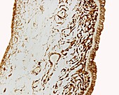
13243896 - Cornea layers, light micrograph
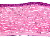
13243889 - Sclera, light micrograph
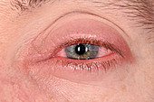
13951631 - Inflamed white of the eye
13736726 - Normal eye and strabismus, illustration
13736721 - Normal eye and eye with cataract, illustration
13613648 - Retina damage from diabetes, fundoscopy
13243905 - Cornea epithelium, light micrograph
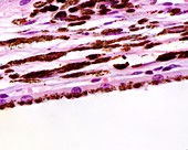
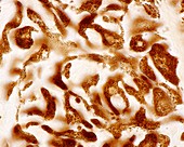
13243900 - Choroid layer, light micrograph
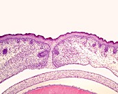
13243886 - Eye development, light micrograph
12987500 - Eye lens, light micrograph
13736719 - Normal eye and eye with cataract, illustration
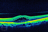
13687017 - Cystoid macular oedema, OCT scan
13636025 - Retina damage from diabetes, illustration
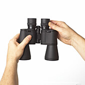
13475045 - Hands adjusting eyepiece of binoculars
13756216 - Human conjunctiva and cornea, light micrograph

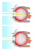
13736717 - Normal eye and eye with glaucoma, illustration
13243903 - Sclera, light micrograph
12991281 - Eye anatomy, illustration
12378553 - Acute glaucoma in a blind eye

13243916 - Cat tapetum lucidum, light micrograph
13243901 - Sclera, light micrograph
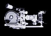
13226573 - Film camera, X-ray
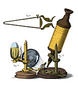
12654348 - Robert Hooke Microscope, 17th Century
13613649 - Retina damage from diabetes, fundoscopy
13377649 - Corrected short-sightedness, illustration
13377643 - Corrected long-sightedness, illustration
13674392 - Liquid gel eye drops
13636058 - Retina damage from diabetes, illustration
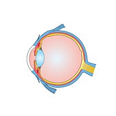
13377637 - Eye anatomy, illustration
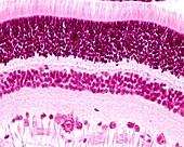
13243919 - Retina layers, light micrograph

13243897 - Cornea, light micrograph
12949097 - Cornea, light micrograph
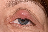
12919357 - Chalazion cyst on an eyelid

12919356 - Bacterial conjunctivitis in HIV patient
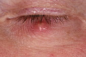
12919355 - Chalazion cyst on an eyelid
12651153 - Cataract Surgery, 2 of 6, Illustration
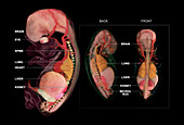
12650244 - Embryo Anatomy, Week 10
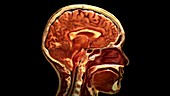
12647808 - Sagittal Section of Head
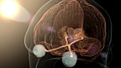
12647806 - Pineal Gland, Sleep Regulation
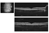
12646750 - Spectral Domain optical coherence tomography of human fovea

12643419 - Anterior uveitis
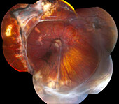
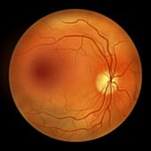
13613664 - Benign eye tumour, fundoscopy
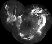
13613651 - Retina damage from diabetes, angiogram
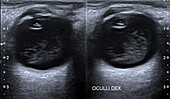
13473917 - Vitreous haemorrhage, ocular ultrasound

13443409 - Microscope and test tubes in a laboratory
13377002 - Rear surface of iris, light micrograph

13376761 - Muscles of the eye, illustration
13243909 - Iris, light micrograph
13243907 - Back of iris, light micrograph
13243898 - Cornea and conjunctiva, light micrograph
page suivante
Oculaire Photos ❘ Science Photo Library