Images
Vidéos
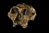
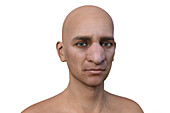
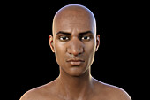
13600855 - Paranthropus aethiopicus, Turkana, Kenya

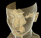
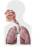
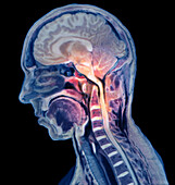
13673785 - Respiratory tract, CT scan
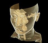
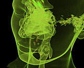
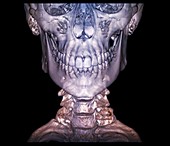
13673919 - Upper respiratory tract, CT scan

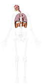
13673773 - Respiratory tract, CT scan
13585352 - Dressing for a nosebleed

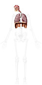
13673786 - Respiratory tract, CT scan
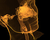
13673775 - Respiratory tract, CT scan
13673921 - Upper respiratory tract, CT scan
13673774 - Respiratory tract, CT scan
13357815 - Concha Bullosa, Illustration
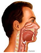
12971288 - Neck anatomy, illustration
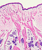
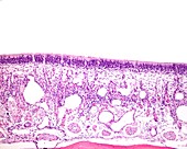
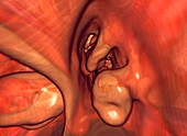
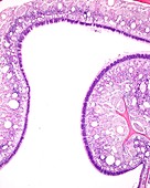
13272208 - Olfactory epithelium section, light micrograph
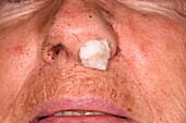
13525415 - Squamous cell carcinoma, LM

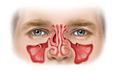
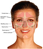
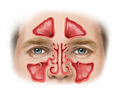
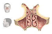
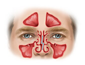
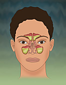
13357814 - Normal Sinuses, Illustration
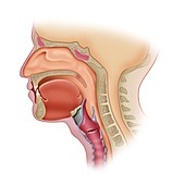
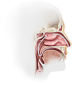
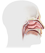
12647808 - Sagittal Section of Head
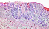
12950643 - Olfactory mucosa of the nasal cavity, light micrograph
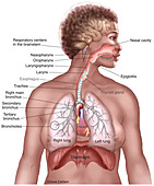
12648600 - Respiratory System Overview (labelled), illustration
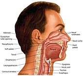
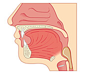
12648569 - Nasal, Oral & Laryngeal Cavities (labelled), illustration
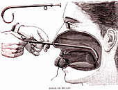
13387778 - Belloc probe, 19th Century illustration
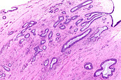
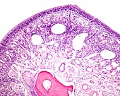
13272210 - Antrochoanal nasal polyp, light micrograph

12950645 - Olfactory mucosa of the nasal cavity, light micrograph
12648598 - Nasal Sinuses (labelled), illustration
12648597 - Nasal Sinuses, illustration
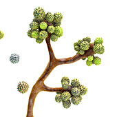
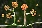
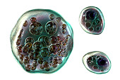
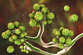
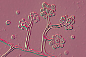
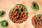
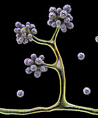
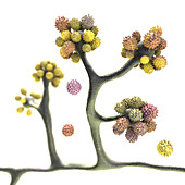
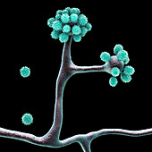
13588696 - Cunninghamella fungi, illustration
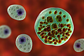
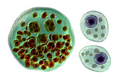
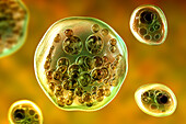
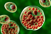
13757666 - Rhinosporidium seeberi parasite, illustration
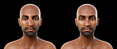
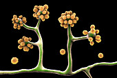
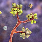
13757024 - Rhinofacial conidiobolomycosis and fungus, illustration
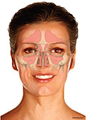
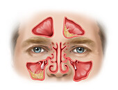
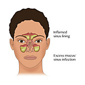
13357676 - Swollen Sinuses, Illustration

13757671 - Rhinosporidium seeberi parasite, illustration
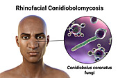
13757019 - Rhinofacial conidiobolomycosis and fungus, illustration
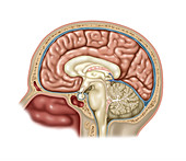
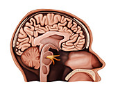
12649627 - Anatomy of Brain, Illustration
12376663 - Nasal cavity, 3D CT scan
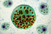
13757664 - Rhinosporidium seeberi parasite, illustration
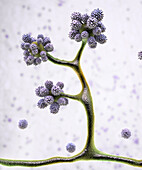
13588708 - Cunninghamella fungi, illustration
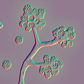
13588697 - Cunninghamella fungi, illustration

13757667 - Rhinosporidium seeberi parasite, illustration
13757022 - Rhinofacial conidiobolomycosis and fungus, illustration
12950644 - Olfactory mucosa of the nasal cavity, light micrograph
13497399 - Swallowing in adult, illustration
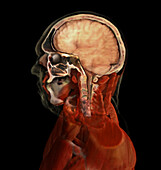
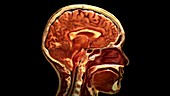
13495240 - Brain and Nasal Cavity, Male Head
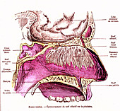
12970457 - Nose anatomy, 19th century
12651183 - Head, Sagittal Section, Illustration
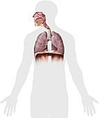
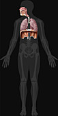
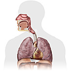
12640542 - Respiratory system, illustration
13588705 - Cunninghamella fungi, illustration
13757672 - Rhinosporidium seeberi parasite, illustration
13757660 - Rhinosporidium seeberi parasite, illustration
13757365 - Nasal cavity, illustration
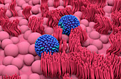
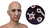
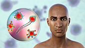
14080088 - Flu viruses on nasal lining, illustration
13757661 - Rhinosporidium seeberi parasite, illustration
13588700 - Cunninghamella fungi, illustration
13357789 - Deviated Septum, Illustration
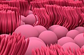
14079265 - Nasal lining, illustration
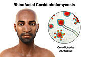
13757015 - Rhinofacial conidiobolomycosis and fungus, illustration
13419972 - Olfactory system, illustration
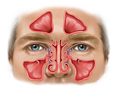
13357760 - Swollen Sinuses with Nasolacrimal Glands, Illustration
12642956 - Nasal cavity, 3D CT scan
12640863 - Nasal Cavity, illustration
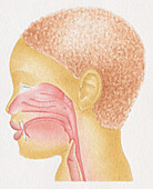
13475587 - Boy's profile outlining the adenoids, illustration
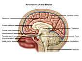
12649626 - Anatomy of Brain, Illustration
12640924 - Respiratory System Upper Organs, illustration
13757013 - Rhinofacial conidiobolomycosis, illustration
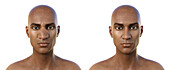
13757012 - Rhinofacial conidiobolomycosis, illustration
13588703 - Cunninghamella fungi, illustration
13357684 - Swollen Infected Sinuses with Polyps, Illustration
12640980 - Organs of the Respiratory System, illustration
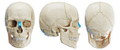
12503894 - Human skull and cervical spine, 3D CT scan
12640327 - Lower respiratory system organs, illustration
12640326 - Organs of the respiratory system, illustration
13757669 - Rhinosporidium seeberi parasite, illustration
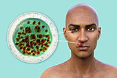
13757026 - Nasal and ocular rhinosporidiosis, illustration
13588704 - Cunninghamella fungi, illustration
13470835 - Nosebleed

12640350 - Upper respiratory system organs, illustration
13757025 - Rhinofacial conidiobolomycosis, illustration
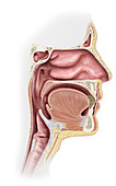
12648566 - Nasal, Oral & Laryngeal Cavities, illustration
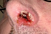
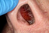
12447502 - Nasal vestibulitis
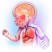
12398306 - Baby's head and chest anatomy, illustration
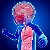
12398256 - Baby's brain and body organs, illustration
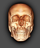
12303241 - Human skull, 3D CT scan
13962383 - Nasal bone, illustration
12640266 - Nasal cavity, illustration
12637880 - Sinusitis, Illustration
13757668 - Rhinosporidium seeberi parasite, illustration
12950648 - Olfactory mucosa of the nasal cavity, light micrograph
12637875 - Sinusitis, Illustration

12446503 - Sinus and ear infections, illustration
14079266 - Nasal lining, illustration
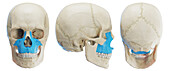
13962382 - Maxilla, illustration
13588707 - Cunninghamella fungi, illustration
13588695 - Cunninghamella fungi, illustration
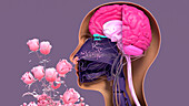
13419970 - Olfactory system, illustration
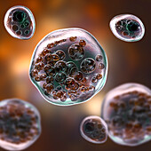
13757663 - Rhinosporidium seeberi parasite, illustration
13757010 - Rhinofacial conidiobolomycosis, illustration
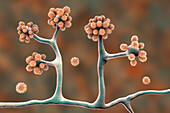
13588706 - Cunninghamella fungi, illustration
13588698 - Cunninghamella fungi, illustration
13588693 - Cunninghamella fungi, illustration
13495258 - Cervical Spine with Rheumatoid Arthritis
12640786 - Respiratory System Organs, illustration
12640732 - Organs of the Upper Body, illustration
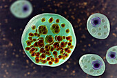
13757665 - Rhinosporidium seeberi parasite, illustration
page suivante
Pathologiste Photos ❘ Science Photo Library