Images
Vidéos

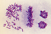
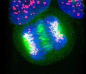
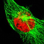
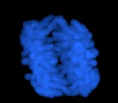
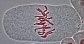
13672564 - HeLa cell in prometaphase, light micrograph
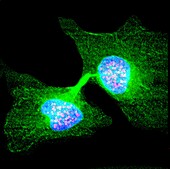
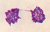
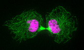
13672550 - Human cell late in cytokinesis, light micrograph
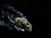
13618781 - Comet C/2020 T2 Palomar
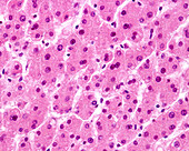
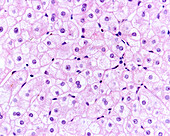
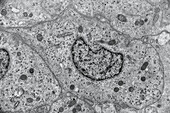
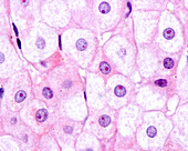
13601238 - Human liver, light micrograph
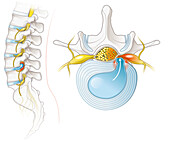
14078629 - Spinal canal stenosis, illustration
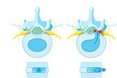
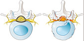
14078589 - Normal disc and herniated disc, illustration
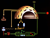
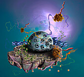
13952310 - Parietal cell, illustration
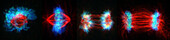
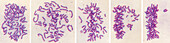
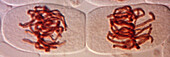
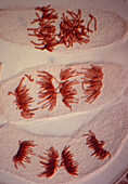
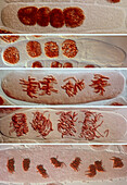
13755099 - Mitosis, light micrograph

13754699 - Mitosis, light micrograph

13754672 - Mitosis, light micrograph
13686216 - Cell anatomy, light micrograph
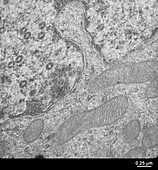
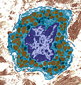
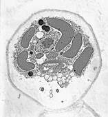
13619631 - Cytomegalovirus, TEM
13601269 - Liver cells, light micrograph
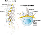
14078580 - Lumbar spine and lumbar vertebra, illustration
13755806 - Mitosis, light micrograph
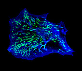
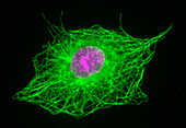
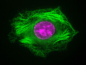
13672567 - Human fibroblast cells, light micrograph
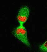
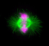
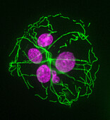
13672536 - Human cell early in cytokinesis, light micrograph
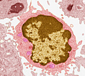
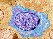
13672259 - Large lymphocyte, TEM
13672046 - Cervical cancer cell, TEM
13632825 - Nucleocytoplasmic transport, illustration
13755812 - Mitosis, light micrograph
13755547 - Mitosis, light micrograph
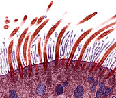
13686361 - Respiratory epithelium, TEM
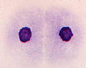
13672544 - Human cell in telophase, light micrograph

13672255 - Large lymphocyte, TEM
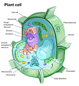
13618818 - Plant cell, illustration
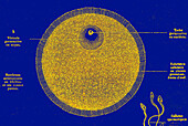
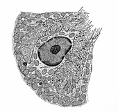
13951717 - Human egg cell, 19th century illustration
13755887 - Mitosis, light micrograph
13755807 - Mitosis, light micrograph

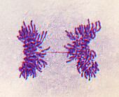
13755805 - Daughter cells after mitosis, light micrograph
13755274 - Daughter cells after mitosis, light micrograph

13742448 - Smooth muscle fibres, light micrograph
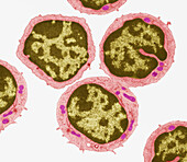
13672261 - Small lymphocytes, TEM
13672044 - Cervical cancer cell, TEM
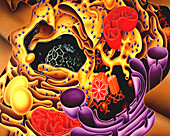
13632782 - Cell interior, illustration
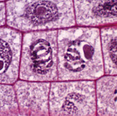
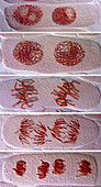
13619593 - Daughter cells, onion root tip, light micrograph
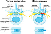
14078590 - Normal disc and herniated disc, illustration
13672555 - Human cell in anaphase, light micrograph
13672547 - Human cell in interphase, light micrograph

13632841 - Cellular protein transport, illustration
13954460 - Mast cell, TEM
13755551 - Mitosis, light micrograph
13754736 - Mitosis in binucleate cells, light micrograph
13672554 - Human cell in telophase, light micrograph
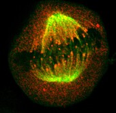
13672552 - Human cell in metaphase, light micrograph
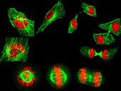
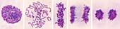
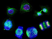
13672549 - Human cells showing the stages of cell division, light micrograph
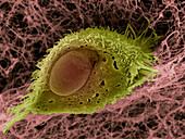
13613362 - Oral squamous cell carcinoma, SEM
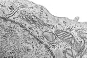
13387605 - Cell structure, TEM
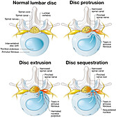
14078594 - Herniated disc progression, illustration
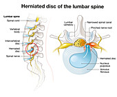
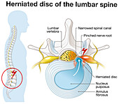
14078566 - Herniated disc of the lumbar spine, illustration
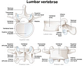
14078563 - Healthy lumbar vertebrae, illustration
13954489 - Mast cell, TEM
13954463 - Trachea, TEM
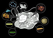
13951731 - Comet Bernardinelli–Bernstein, illustration
13755097 - Mitosis, light micrograph
13754706 - Mitosis, light micrograph
13672557 - Two human cells in interphase, light micrograph
13672533 - Human cell in anaphase, light micrograph
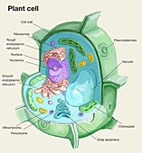
13618820 - Plant cell, illustration
13601267 - Liver cells, light micrograph
14078567 - Herniated disc of the lumbar spine, illustration
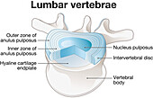
14078561 - Healthy lumbar vertebrae and intervertebral disc, illustration
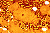
13756209 - Dorsal root ganglion, light micrograph
13755895 - Interphase cell treated with Taxol, light micrograph
13755889 - Mitosis, light micrograph
13755804 - Mitosis, light micrograph
13754738 - Mitosis in binucleate cells, light micrograph
13672566 - Human fibroblast cells, light micrograph
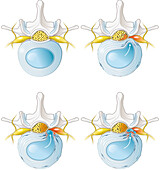
14078593 - Herniated disc progression, illustration
13754741 - Mitosis in tetranucleate cells, light micrograph
13672556 - Human cell in early prophase, light micrograph
13647381 - Protein synthesis, illustration
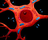
13632840 - Nerve cell, illustration
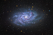
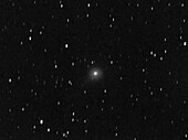
13297448 - Triangulum galaxy (M33)
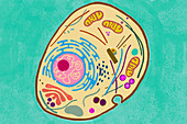
13951826 - Animal cell structure, illustration
13755896 - Interphase cell treated with Colchicine, light micrograph
13755891 - Cytokinesis, light micrograph

13754742 - Mitosis in tetranucleate cells, light micrograph

13733278 - Animal cell structure, illustration

13673736 - Carbon-12, atomic structure

13672561 - Human cell in metaphase, light micrograph
13672553 - Stages of cell division, light micrograph
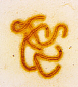
13672532 - Human chromosomes in early anaphase, light micrograph
13672256 - Small lymphocytes, TEM

13619595 - Cytokinesis in onion root tip cell, light micrograph
14078565 - Herniated disc of the lumbar spine, illustration
13954472 - Somatotroph, TEM
13755886 - Mitosis, light micrograph
13754700 - Mitosis, light micrograph
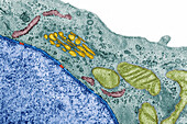
13619461 - Cell organelles, TEM
13601058 - Animal cell, illustration
13496605 - Myelocyte, Basophil, LM

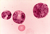
13496554 - Promyelocyte, LM

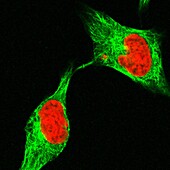
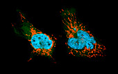
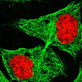
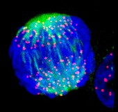
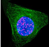
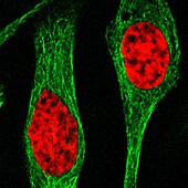
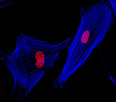
13473900 - Fibroblast, fluorescent micrograph
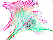
13473886 - Fibroblast, fluorescent micrograph
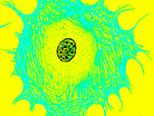
13473882 - Fibroblast, fluorescent micrograph
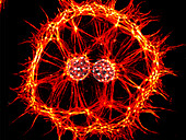
13473875 - Fibroblast, fluorescent micrograph
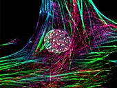
13473797 - Fibroblast, fluorescent micrograph
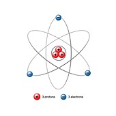
13377467 - Lithium atom, illustration
page suivante
Nucleus Photos ❘ Science Photo Library