Human cell in telophase, light micrograph
Numéro d’image : 13672554
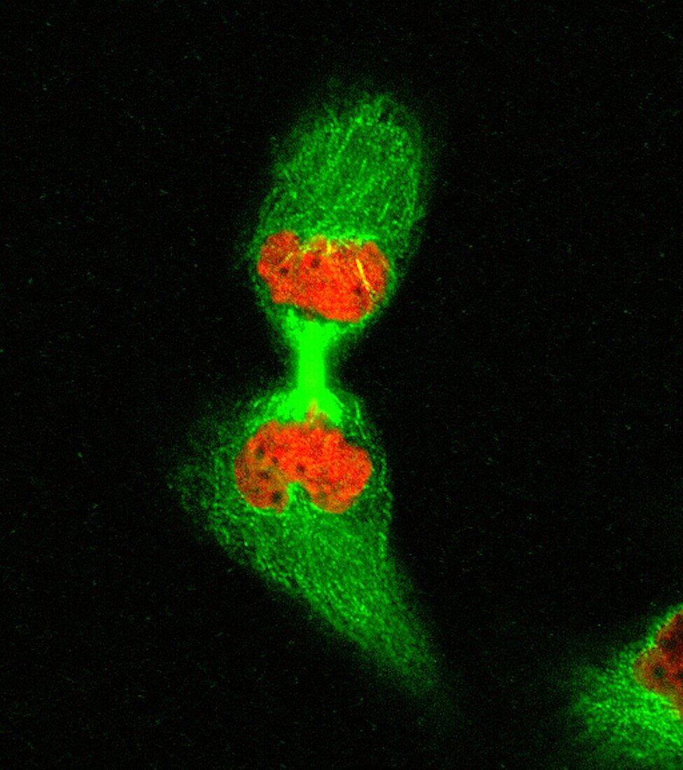
| HeLa cell in telophase, fluorescence light micrograph. Chromatin is stained red and the microtubules forming the spindle are stained green. The chromosomes have separated and decondensed, and the midbody has formed. | |
| Licence : | Droits gérés |
| Crédit: | Science Photo Library / DR MATTHEW DANIELS |
| Taille de l’image : | 3050 px × 3439 px |
| Model Release : | Non requis |
| Property Release : | Non requis |
| Restrictions : | - |
Prix pour cette image À partir de 45 €
Produit vendu
(Calendrier, Carte postale, Carte de vœux, Impression sur textile, Packaging etc)
À partir de 45 €
Usage commercial
(Affichage, Annonce presse, Annonce TV, Carte, Digital - hors rés. sociaux, Digital - rés. sociaux etc)
À partir de 45 €
Éditorial
(Digital, Journal, Livre, Livre pratique, Magazine, Télévision etc)
À partir de 60 €
Usage non-commercial
(Digital - hors rés. sociaux, Digital - rés. sociaux etc)
À partir de 120 €
Mots clés
- arrière plan noir,
- arrière-plan noir,
- aucun,
- axe,
- biologie,
- biologie cellulaire,
- biologique,
- broche,
- chromatine,
- chromosomes,
- corps central,
- cytologie,
- cytologique,
- division cellulaire,
- division nucléaire,
- fluorescence,
- fluorescent,
- fond noir,
- génétique,
- microscope,
- microscope optique,
- microscopie,
- microscopie optique,
- microtubules,
- mitose,
- noyau,
- nucleus,
- personne,
- telophase,
- tige