Images
Vidéos
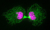
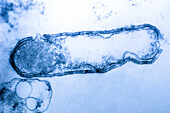
13755891 - Cytokinesis, light micrograph
13754875 - Premature chromosome condensation, light micrograph

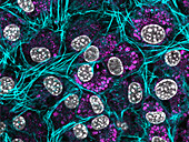
13754742 - Mitosis in tetranucleate cells, light micrograph
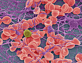
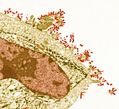
13743102 - Blood clot, SEM
13743099 - PICC line internal surface, SEM

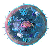
13733278 - Animal cell structure, illustration
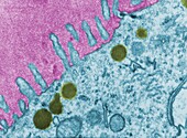
13673233 - Flu, TEM

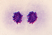
13672561 - Human cell in metaphase, light micrograph
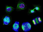
13672553 - Stages of cell division, light micrograph
13672532 - Human chromosomes in early anaphase, light micrograph
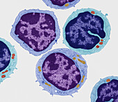
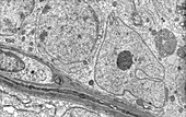
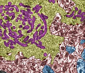
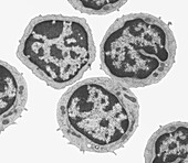
13672256 - Small lymphocytes, TEM
13633134 - Human induced pluripotent cell
13613421 - Nerve, TEM
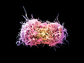
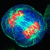
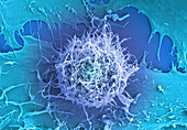
13613369 - Lung cancer cells dividing, SEM
13525474 - Gap junction, TEM

13954469 - Dividing cancer cell, TEM
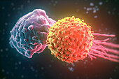
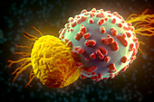
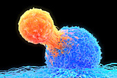
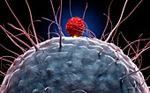
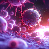
13765645 - T cell attacking cancer cell, illustration
13755936 - Spermatid chromatoid body, TEM
13755886 - Mitosis, light micrograph

13754878 - Condensed chromosomes, light micrograph
13754700 - Mitosis, light micrograph
13672828 - Human 80s ribosome, illustration
13672534 - Human cell showing nucleus and tubulin, light micrograph
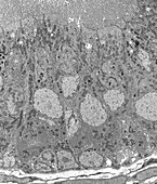
13634182 - Testis, SEM

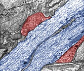
13620785 - Microvilli, TEM
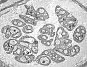
13613428 - Epididymis, TEM
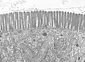
13613407 - Microvilli, TEM
13613398 - Sertoli cell, TEM
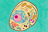
13601058 - Animal cell, illustration

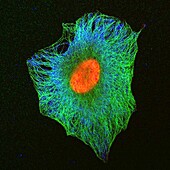
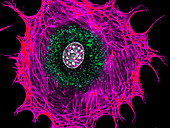
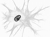
13473900 - Fibroblast, fluorescent micrograph
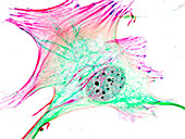
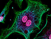
13473886 - Fibroblast, fluorescent micrograph
13473882 - Fibroblast, fluorescent micrograph
13473875 - Fibroblast, fluorescent micrograph
13473797 - Fibroblast, fluorescent micrograph

13405953 - Microtubule, computer model
13386718 - T cells attacking cancer cells, illustration
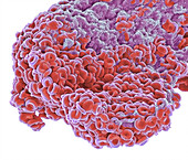
13360083 - Megakaryocytes and Erythrocytes, EM
13356875 - Ovarian fibroma, TEM

13356054 - Breast cancer, TEM
13272764 - Cell membrane, illustration
13272291 - Cell cycle, illustration
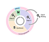
13272289 - Cell cycle, illustration
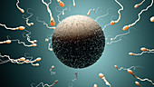
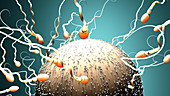
13223389 - Fertilisation, illustration
12987538 - Mitosis, light micrograph
12961643 - Natural killer T cell attacking cancer cell,illustration
13765638 - T cell attacking cancer cell, illustration
13755890 - Mitosis, light micrograph
13754880 - Mitosis, light micrograph

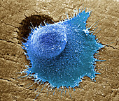
13743100 - Cervial cancer cell, SEM.
13733279 - Animal cell structure, illustration
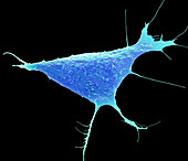
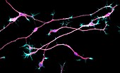
13686222 - Stem cell-derived neurons, light micrograph
13673240 - Flu, TEM

13613417 - Dividing cell, TEM
13525552 - Rabies virus, TEM
13473898 - Fibroblast, fluorescent micrograph
13473887 - Fibroblast, fluorescent micrograph

13473809 - Fibroblast, fluorescent micrograph
13473127 - Axodendritic synapses, TEM
13473125 - Cerebellar glomerulus, TEM
13473113 - Oocyte, TEM

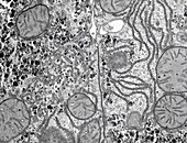
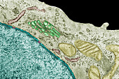
13473108 - Endoplasmic reticulum, TEM
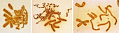
13357514 - Transmission Electron Microscopy (TEM) of Clostridium botulinum Bacteria
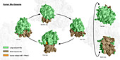
13244504 - Mitochondrial changes in cell reprogramming, illustration
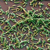
13243986 - Aeromonas hydrophila bacteria, SEM
13225504 - T-cell attaching to cancer cell, illustration
13223388 - Fertilisation, illustration

12987539 - Mitosis, light micrograph
12961625 - Dendritic cell and T cell,illustration

12961624 - Dendritic cell and T cell,illustration
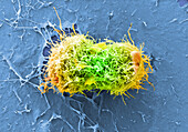
13838889 - Cervical cancer cells dividing, SEM
13755107 - Mitosis, light micrograph
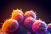
13686468 - Bone Cancer, SEM
13672563 - Human cell in anaphase, light micrograph
13672260 - Small lymphocytes, TEM
13672043 - Cervical cancer cell, TEM
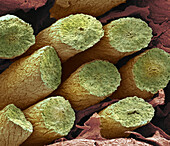
13633210 - Eye lens fibres, SEM
13633174 - Human induced pluripotent cell.
13599623 - IVF embryo, light micrograph
13473854 - Fibroblast, fluorescent micrograph
13473843 - Fibroblasts, fluorescent micrograph

13473834 - Fibroblast, fluorescent micrograph
13473813 - Fibroblast, fluorescent micrograph
13473802 - Fibroblasts, fluorescent micrograph

13473112 - Oocyte, TEM
13387801 - Brain hippocampus neurons, fluorescence light micrograph
13386713 - T cell attacking cancer cell
13361094 - Basophils, SEM

13356877 - Platelet, TEM
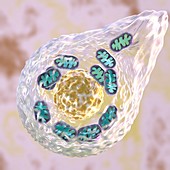
13244502 - Mitochondrial changes in cell reprogramming, illustration

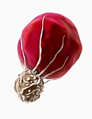
13223383 - Sperm, illustration

12961626 - Dendritic cell and T cell,illustration
12961623 - Dendritic cells,illustration
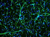
12961067 - Neurons from stem cells,fluorescence light micrograph
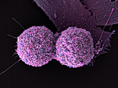
13952273 - Cervical cancer cells, SEM
13838886 - Cervical cancer cells dividing, SEM
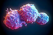
13765625 - Cancer cells, illustration
13765623 - Cancer cells, illustration
13765613 - Cancer cells, illustration

13755290 - Mitosis in topoisomerase II inhibited cell, light micrograph
page suivante
Cytologique Photos ❘ Science Photo Library