Fibroblast, fluorescent micrograph
Numéro d’image : 13473834
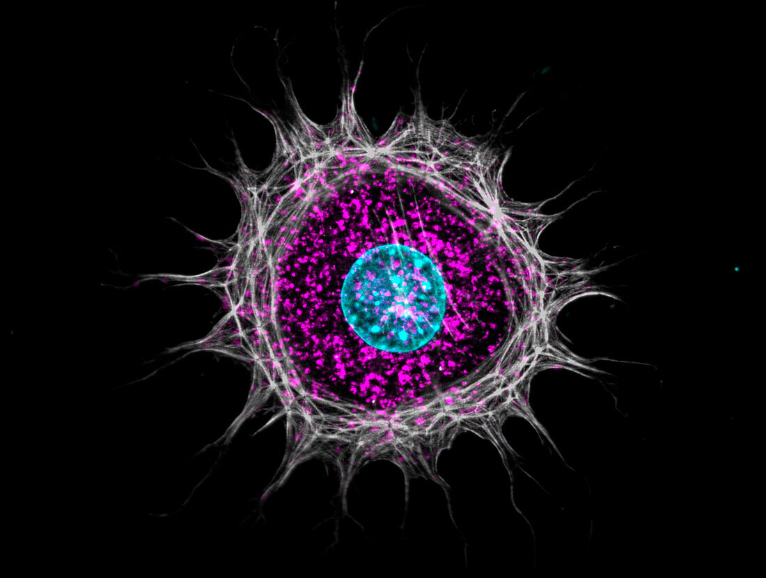
| Immunofluorescence micrograph of a murine fibroblast stained with an actin cytoskeleton probe (white), anti-LAMP-1 (lysosomal-associated membrane protein 1) antibody (purple), and nuclear DAPI (light blue). Focal planes were achieved with an optical apotome. | |
| Licence : | Droits gérés |
| Crédit: | Science Photo Library / JACOB C. ZBINDEN |
| Taille de l’image : | 6808 px × 5134 px |
| Model Release : | Non requis |
| Property Release : | Non requis |
| Restrictions : | - |
Prix pour cette image À partir de 45 €
Produit vendu
(Calendrier, Carte postale, Carte de vœux, Impression sur textile, Packaging etc)
À partir de 45 €
Usage commercial
(Affichage, Annonce presse, Annonce TV, Carte, Digital - hors rés. sociaux, Digital - rés. sociaux etc)
À partir de 45 €
Éditorial
(Digital, Journal, Livre, Livre pratique, Magazine, Télévision etc)
À partir de 60 €
Usage non-commercial
(Digital - hors rés. sociaux, Digital - rés. sociaux etc)
À partir de 120 €
Mots clés
- actine,
- aucun,
- biologie,
- biologique,
- cellulaire,
- cellule,
- cellules,
- cytologie,
- cytologique,
- cytosquelette,
- cytosquelettique,
- DAPI,
- fibroblaste,
- fluorescence,
- fluorescent,
- immunofluorescence,
- lampe,
- mammifère,
- microscope,
- microscope optique,
- microscopie,
- microscopie optique,
- murin,
- murine,
- noyau,
- nucleus,
- personne,
- protéine,
- sali,
- souillé,
- souris,
- tache,
- taché