Images
Vidéos
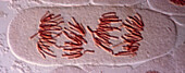
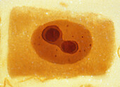
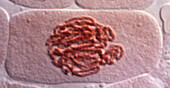
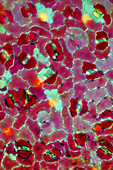
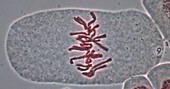
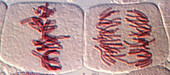
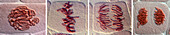
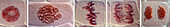
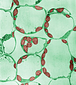
13754734 - Mitosis in binucleate cells, light micrograph

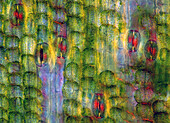
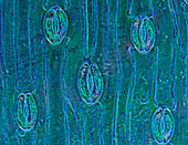
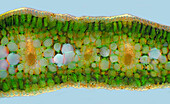
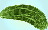
13634300 - Water milfoil, light micrograph
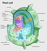
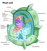
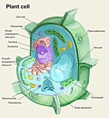
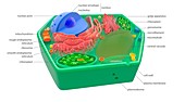
13618819 - Plant cell, illustration
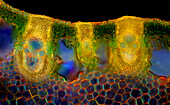
13954159 - Stomata in hyacinth (Hyacinthus sp.) leaf, light micrograph
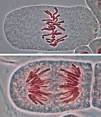
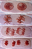
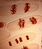
13754701 - Mitosis, light micrograph
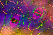
13754752 - Segregated nucleolus, light micrograph
13954538 - Stomata in tulip leaf epidermis, light micrograph
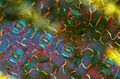
13954442 - Stomata in spath leaf epidermis, light micrograph
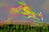
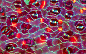
13633940 - Calcium oxalate crystals in field scabious, light micrograph
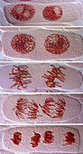
13754707 - Mitosis, light micrograph
13954448 - Stomata in spath leaf epidermis, light micrograph
13954393 - Stomata in croton leaf, light micrograph

13754699 - Mitosis, light micrograph

13754672 - Mitosis, light micrograph
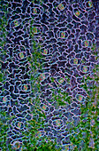
13634298 - Calcium oxalate crystals in water milfoil, light micrograph
13954388 - Stomata in croton leaf, light micrograph
13954179 - Hyacinth (Hyacinthus sp.) leaf, light micrograph
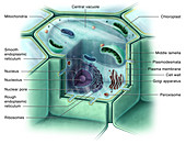
13618818 - Plant cell, illustration
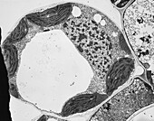
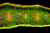
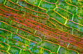
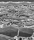
13618624 - Timothy grass mesophyll cell, TEM
13954447 - Stomata in spath leaf epidermis, light micrograph
13954441 - Stomata in spath leaf epidermis, light micrograph
13954180 - Hyacinth (Hyacinthus sp.) leaf, light micrograph
13754736 - Mitosis in binucleate cells, light micrograph
13954161 - Stomata in hyacinth (Hyacinthus sp.) leaf, light micrograph
13754706 - Mitosis, light micrograph
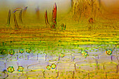
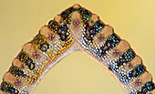
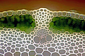
13633783 - Senecio sp. sclerenchyma and collenchyma, light micrograph
13618820 - Plant cell, illustration
13954444 - Stomata in spath leaf epidermis, light micrograph
13754738 - Mitosis in binucleate cells, light micrograph
13754741 - Mitosis in tetranucleate cells, light micrograph
13954160 - Stomata in hyacinth (Hyacinthus sp.) leaf, light micrograph

13754742 - Mitosis in tetranucleate cells, light micrograph
13754700 - Mitosis, light micrograph
13954163 - Raphides from hyacinth leaf, light micrograph
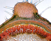
13634108 - Grass stalk, light micrograph
13504001 - Grass leaf, polarised light micrograph

13754705 - Mitosis, light micrograph
13754702 - Mitosis, light micrograph
13754737 - Mitosis in binucleate cells, light micrograph
13503980 - Canadian pondweed (Elodea canadensis), light micrograph
13954392 - Stomata in croton leaf, light micrograph
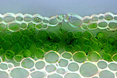
13634299 - Water milfoil stalk, light micrograph
13954446 - Stomata in spath leaf epidermis, light micrograph
13954443 - Stomata in spath leaf epidermis, light micrograph
13954394 - Stomata in croton leaf, light micrograph
13503981 - Canadian pondweed (Elodea canadensis), light micrograph
13954537 - Stomata in tulip leaf epidermis, light micrograph
13954445 - Stomata in spath leaf epidermis, light micrograph
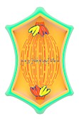
13754753 - Nucleolar cycle, light micrograph
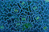
13634308 - Aquatic plant with cyanobacteria and algae, light micrograph
13634293 - Water milfoil stalk, light micrograph
13754673 - Mitosis, light micrograph
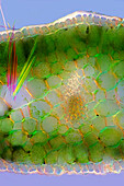
13503519 - Spring vetchling leaf stalk, light micrograph
13754704 - Mitosis, light micrograph
13954183 - Hyacinth leaf with raphides, light micrograph
13954178 - Hyacinth (Hyacinthus sp.) leaf, light micrograph
13754745 - Mitosis in binucleate cells, light micrograph
13754703 - Mitosis, light micrograph
13504000 - Grass stalk tissues, polarised light micrograph
13954164 - Hyacinth leaf with raphides, light micrograph
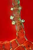
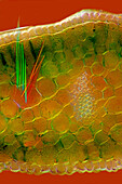
13634017 - Field scabious (Knautia arvensis) stoma, light micrograph
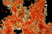
13504010 - Haematococcus pluvialis algae, light micrograph
13503717 - Lily stalk, polarized light micrograph

13506080 - Onion root cell, TEM
12249571 - Plant cell, illustration
13504023 - Mentha stalk, polarised light micrograph

13495304 - Duckweed root cells, TEM
13503989 - Elodea canadensis stalk, polarised light micrograph
13504003 - Vascuclar bundles in grass leaf, polarised light micrograph
12606037 - Corn Root Tip Cell, TEM
13504012 - Haematococcus pluvialis, light micrograph
12635799 - Osmosis in Plant Cells, Illustration

12635728 - Plant Cell, Illustration

12349392 - Wanda K. Farr, US botanist
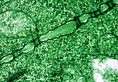
13495298 - Spinach Leaf Mesophyll, TEM
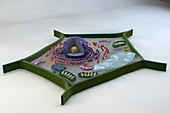
12639248 - Plant Cell, Illustration
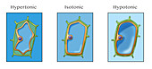
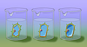
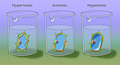
12637038 - Osmosis in Plant and Animal Cells, Illustration
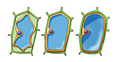
12635802 - Osmosis in Plant Cells, Illustration
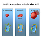
12637037 - Osmosis in Plant and Animal Cells, Illustration

12635800 - Osmosis in Plant Cells, Illustration
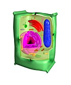
12644437 - Plant cell, illustration
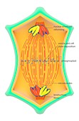
11703763 - Cytokinesis in plant cells,illustration
12047081 - Corn Leaf Chloroplast,TEM
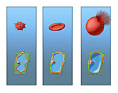
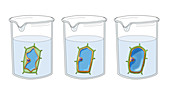
12635797 - Osmosis in Plant Cells, Illustration
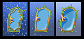
12635801 - Osmosis in Plant Cells, Illustration
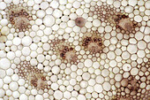
12378184 - Apple fruit section, skin to parenchyma, SEM
11703637 - Plant cell,illustration
12635727 - Plant Cell, Illustration
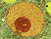
12016418 - Spinach Leaf Mesophyll (TEM)
11703764 - Cytokinesis in plant cells,illustration
12635798 - Osmosis in Plant Cells, Illustration
12635796 - Osmosis in Plant Cells, Illustration
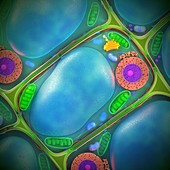
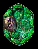
13437544 - Plant cell structure, illustration
11701465 - Plasmodesmata plant cell junctions,TEM
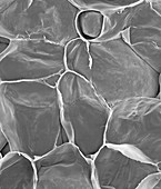
12378183 - Apple parenchyma cells (Malus domestica), SEM
12047493 - Magnolia Stem,LM
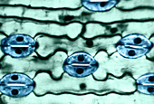
12047486 - Leaf Epidermis,LM
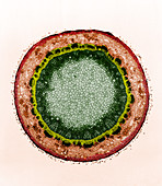
12047170 - Thale Cress,Root Tip Cell Nucleus,TEM

12046707 - Iodine Stained Onion Cells
page suivante
Cellule végétale Photos ❘ Science Photo Library