Eye socket anatomy, illustration
Numéro d’image : 12969959
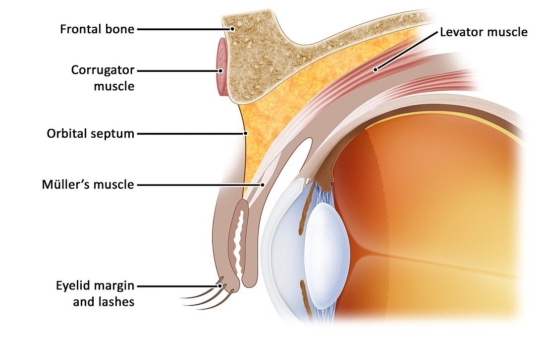
| Eye socket anatomy, illustration. This is a sagittal view, with the front of the eye at left. The structures and tissues labelled here are the frontal bone, the corrugator muscle, the levator muscle of the eyelid, the orbital septum, Mueller's muscle (the superior tarsal muscle, another of the eyelid muscles), and the eyelid margin and eyelashes. The lens of the eye and its cornea is also shown. | |
| Licence : | Droits gérés |
| Crédit: | Science Photo Library / De Angelis, Maurizio |
| Taille de l’image : | 4000 px × 2502 px |
| Model Release : | Non requis |
| Property Release : | Non requis |
| Restrictions : | - |
Prix pour cette image À partir de 45 €
Produit vendu
(Calendrier, Carte postale, Carte de vœux, Impression sur textile, Packaging etc)
À partir de 45 €
Usage commercial
(Affichage, Annonce presse, Annonce TV, Carte, Digital - hors rés. sociaux, Digital - rés. sociaux etc)
À partir de 45 €
Éditorial
(Digital, Journal, Livre, Livre pratique, Magazine, Télévision etc)
À partir de 60 €
Usage non-commercial
(Digital - hors rés. sociaux, Digital - rés. sociaux etc)
À partir de 120 €
Mots clés
- anatomie,
- anatomique,
- arrière plan blanc,
- arrière-plan blanc,
- aucun,
- biologie,
- biologique,
- catégorie,
- cils,
- cornea,
- cornée,
- corps humain,
- coupe,
- divisé,
- en bonne santé,
- étiqueté,
- étiquette,
- étiquettes,
- fond blanc,
- illustration,
- label,
- lentille,
- marqué,
- muscle,
- normal,
- oculaire,
- oeil,
- oeuvre,
- ophtalmologie,
- os,
- partie,
- paupière,
- personne,
- prise,
- sagittal,
- sagittale,
- sain,
- section,
- sourcils,
- texte