Rods and cones in retina
Numéro d’image : 12070707
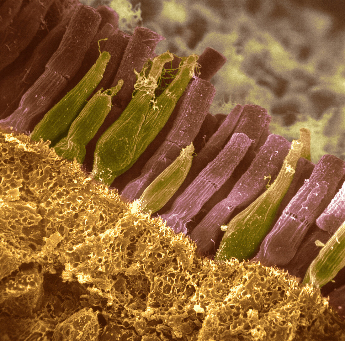
| colourized scanning electron micrograph (SEM) of rods and cones,showing the structure of the eye retina. The retina is a thin tissue layer on the inner eye responsible for sight. Light strikes from the top. At top are nerve fibers which combine to form the optic nerve to the brain. Nerve fibers have round cell bodies with branching dendrites. Rods (brown) are long nerve cells which respond to dim light,enabling images to be detected. Cones (olive coloured) are shorter cone-like cells which detect colour. Rods and cones pass visual signals through the optic nerve to the brain. Pigment cells block light from passing further. Magnification: unknown. Enhancement of 9B9376 | |
| Licence : | Droits gérés |
| Crédit: | Science Photo Library / Omikron |
| Taille de l’image : | 3300 px × 3265 px |
| Model Release : | Non requis |
| Property Release : | Non requis |
| Restrictions : |
|
Prix pour cette image À partir de 45 €
Produit vendu
(Calendrier, Carte postale, Carte de vœux, Impression sur textile, Packaging etc)
À partir de 45 €
Usage commercial
(Affichage, Annonce presse, Annonce TV, Carte, Digital - hors rés. sociaux, Digital - rés. sociaux etc)
À partir de 45 €
Éditorial
(Digital, Journal, Livre, Livre pratique, Magazine, Télévision etc)
À partir de 60 €
Usage non-commercial
(Digital - hors rés. sociaux, Digital - rés. sociaux etc)
À partir de 120 €
Mots clés
- anatomie,
- bâtonnet,
- bâtonnets,
- cône,
- cônes,
- cornea,
- cornée,
- corps,
- élève,
- fibre nevreuse,
- fibres nerveuses,
- globe oculaire,
- humain,
- iris,
- lentille,
- M.E.B.,
- MEB,
- micrographe,
- micrographeà balayage électronique,
- micrographie,
- microscope,
- microscope à balayage électronique,
- microscope électronique à balayage,
- microscopie,
- nerf optique,
- oeil,
- retina,
- rétine,
- sens,
- synapse,
- tige,
- tiges,
- vision,
- visuel,
- vue