Juvenile Nasopharyngeal Angiofibroma
Numéro d’image : 12006578
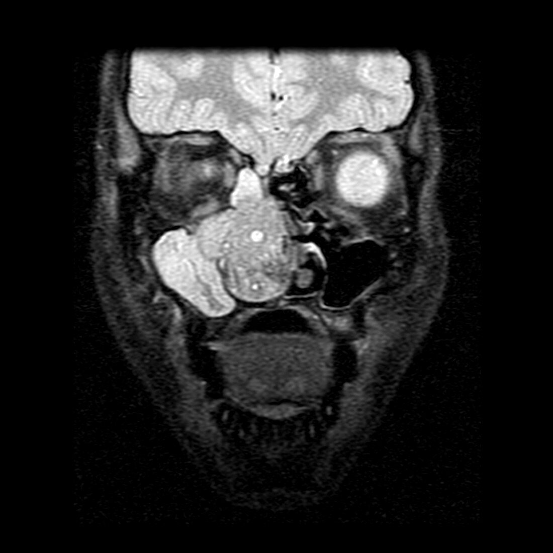
| This coronal (frontal) T2 weighted MRI image of the head in an adolescent male demonstrates a large irregular mass in the right (on your left) nasal cavity with obstruction of drainage of the right maxillary sinus and right ethmoid air cells which are completely filled with mucus/fluid. The mass in the right nasal cavity represents a benign but invasive tumor called a juvenile nasopharyngeal angiofibroma which typically occur in adolescent males | |
| Licence : | Droits gérés |
| Crédit: | Science Photo Library / Living Art Enterprises |
| Taille de l’image : | 6000 px × 6000 px |
| Model Release : | Non requis |
| Property Release : | Non requis |
| Restrictions : |
|
Prix pour cette image À partir de 45 €
Produit vendu
(Calendrier, Carte postale, Carte de vœux, Impression sur textile, Packaging etc)
À partir de 45 €
Usage commercial
(Affichage, Annonce presse, Annonce TV, Carte, Digital - hors rés. sociaux, Digital - rés. sociaux etc)
À partir de 45 €
Éditorial
(Digital, Journal, Livre, Livre pratique, Magazine, Télévision etc)
À partir de 60 €
Usage non-commercial
(Digital - hors rés. sociaux, Digital - rés. sociaux etc)
À partir de 120 €
Mots clés
- angiofibrome nasopharyngé juvénile,
- cavité nasale,
- epistaxis,
- I.R.M.,
- Image à Résonnance Magnétique de la tête,
- imagerie par résonance magnétique,
- imagerie par résonnance magnétique,
- IRM,
- IRM de la tête,
- JNA,
- masse nasale,
- monochrome,
- N/B,
- nasal,
- NB,
- néoplasme,
- nez,
- noir blanc,
- noir et blanc,
- pathologiste,
- pédiatrique,
- sinus,
- sinus paranasal,
- sinus paranasaux,
- sinus parnasal,
- tête,
- tumeur