Knee anatomy,artwork
Numéro d’image : 11626803
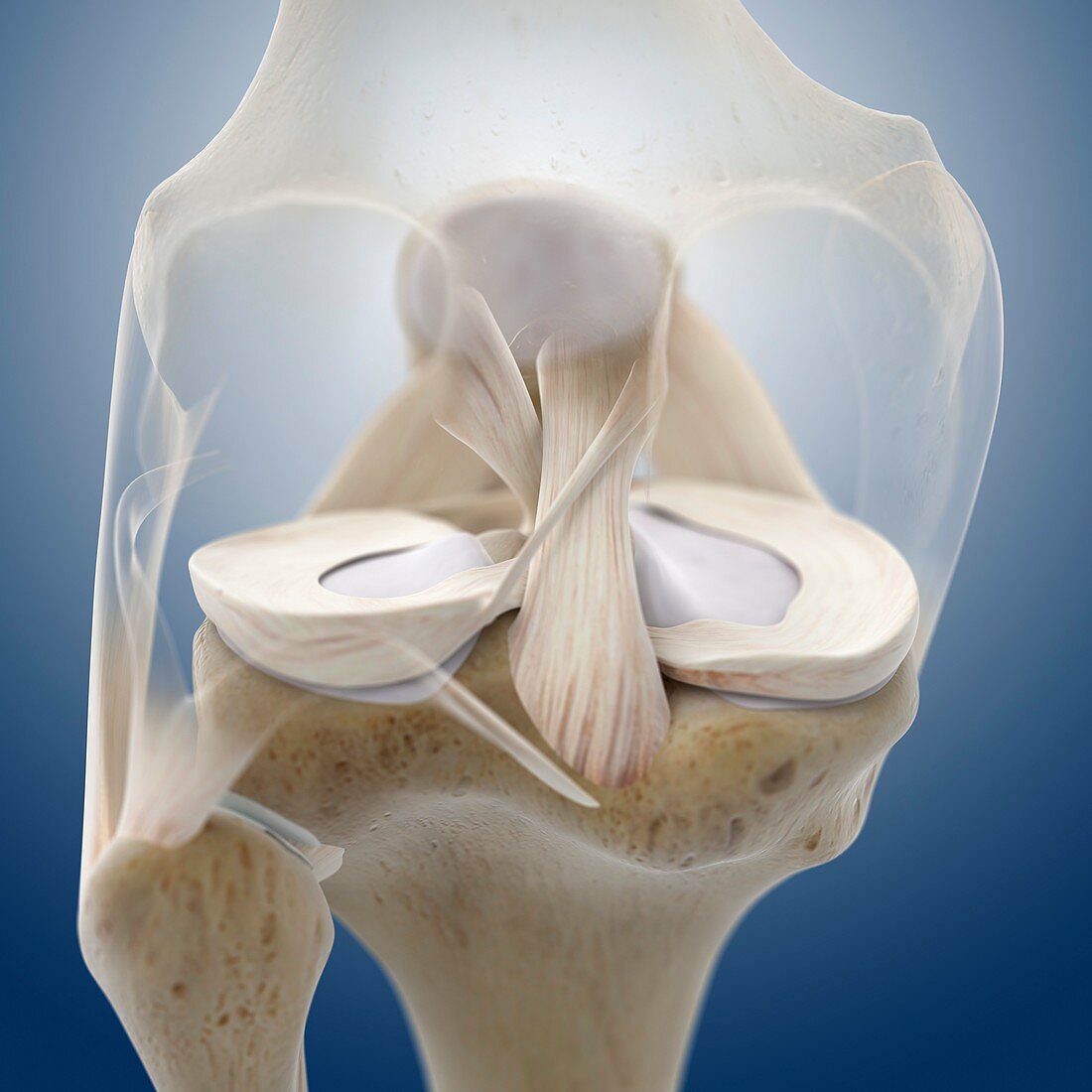
| Knee anatomy,computer artwork. The knee joint is formed by the articulation of the femur (thigh bone,opaque,top) with tibia (shin bone,bottom centre). The smaller fibula (bottom left) is also seen. Articular cartilage (white),which protects the bone surface from wear and tear,is seen on the head of the tibia. Partially covering it are the menisci (cream crescent-shaped),which act as shock absorbers. At centre is the posterior cruciate ligament,a fibrous band of connective tissue that connects the femur and tibia and limits backwards movement of the joint | |
| Licence : | Droits gérés |
| Crédit: | Science Photo Library / Springer Medizin |
| Taille de l’image : | 4180 px × 4180 px |
| Model Release : | Non requis |
| Property Release : | Non requis |
| Restrictions : | - |
Prix pour cette image À partir de 45 €
Produit vendu
(Calendrier, Carte postale, Carte de vœux, Impression sur textile, Packaging etc)
À partir de 45 €
Usage commercial
(Affichage, Annonce presse, Annonce TV, Carte, Digital - hors rés. sociaux, Digital - rés. sociaux etc)
À partir de 45 €
Éditorial
(Digital, Journal, Livre, Livre pratique, Magazine, Télévision etc)
À partir de 60 €
Usage non-commercial
(Digital - hors rés. sociaux, Digital - rés. sociaux etc)
À partir de 120 €
Mots clés
- anatomie,
- anatomique,
- arrière,
- articulation,
- biologie,
- biologique,
- cartilage,
- cartilage hyalin,
- corps humain,
- cuisse,
- de retour,
- dos,
- en bonne santé,
- fémoral,
- fémur,
- fibula,
- genou,
- illustration,
- jambe,
- LCP,
- ligament croisé postérieur,
- ligament croisé postéro-interne,
- membre inférieur,
- menisces,
- menisci,
- ménisque,
- normal,
- oeuvre,
- os,
- os de la cuisse,
- péroné,
- postérieur,
- sain,
- squelette,
- système squelettique,
- tibia,
- tissu conjonctif