Images
Vidéos
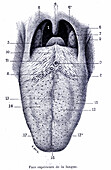
13736195 - Tongue anatomy, illustration
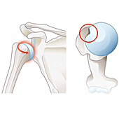
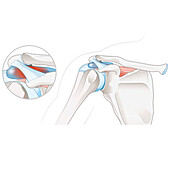
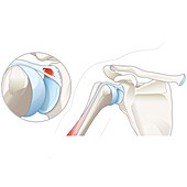
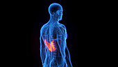
14078633 - Subacromial bursitis of the shoulder, illustration
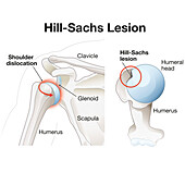
14078575 - Hill-Sachs lesion of the shoulder, illustration

13950597 - Human tongue

13736157 - Anatomy of ischemic stroke, illustration
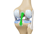
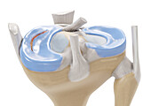
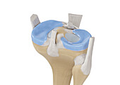
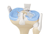
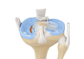
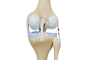
14078950 - Posterior cruciate ligament, illustration
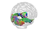
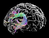
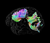
13950870 - White matter fibres, DTI MRI scan
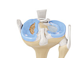
14078917 - Zones blood flow meniscus, illustration
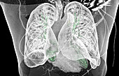
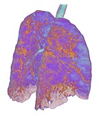
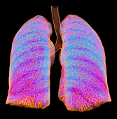
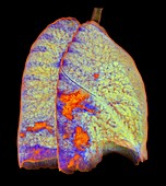
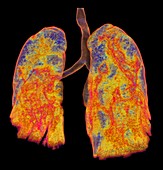
13952114 - Healthy lungs, CT scan
14078934 - Longitudinal tear meniscus, illustration

14077843 - Human forearm muscles, 1844 illustration
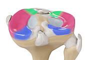
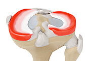
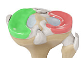
14078945 - Meniscus regions, illustration
14078635 - Subacromial bursitis of the shoulder, illustration
14078920 - Bucket handle tear meniscus, illustration
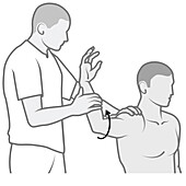
14078610 - Shoulder examination, illustration
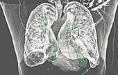
13952116 - Healthy lungs, CT scan
14078941 - Menisci lateral posterior, illustration

13950952 - Superior and inferior cerebellar peduncles, DTI MRI scan
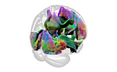
13950872 - White matter fibres, DTI MRI scan

14078634 - Subacromial bursitis of the shoulder, illustration

13435962 - Female leg musculature, illustration
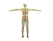
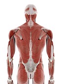
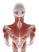
13435550 - Muscular system, illustration

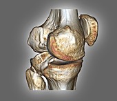
13243024 - Healthy knee, CT scan
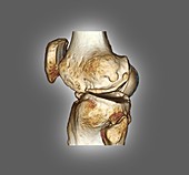
13243023 - Healthy knee, CT scan
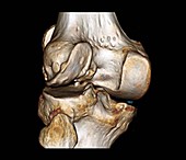
13243020 - Healthy knee, CT scan
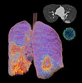
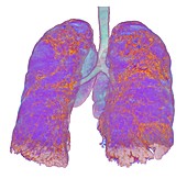
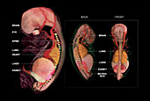
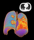
13218637 - Lungs affected by Covid-19, CT scans and model
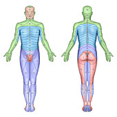
14077605 - Dermatome skin sensory areas, illustration
13224585 - Muscles of the back, illustration
14078952 - Radial meniscus tear, illustration

13471408 - Post pelvic exenteration surgery
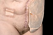
13471328 - Post pelvic exenteration surgery

13435913 - Kidney anatomy, illustration
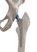
13435814 - Hip replacement, artwork
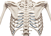
13435230 - Rib cage, illustration

13950875 - White matter fibres, DTI MRI scan

13443466 - Thyroid gland, illustration
13436093 - Lymphatic system, illustration
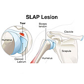
14078626 - SLAP lesion of the shoulder, illustration
13950914 - Arcuate fasciculus, DTI MRI scan
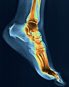
13452941 - Heel pain, X-ray
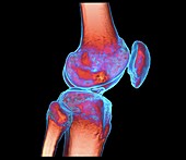
13243027 - Healthy knee, CT scan

13218638 - Lungs affected by Covid-19 atypical pneumonia, CT scans
13218170 - Lungs affected by Covid-19 atypical pneumonia, 3d CT scan

13950951 - Inferior cerebellar peduncle, DTI MRI scan
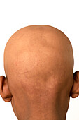
13444554 - Alopecia totalis

13435861 - Thoracic spine, illustration
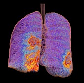
13272744 - Aeration pattern in healthy lungs, 3D CT scan
13224580 - Muscles of the back, illustration
13218168 - Lungs affected by Covid-19 atypical pneumonia, 3d CT scan
13218159 - Lungs affected by Covid-19 atypical pneumonia, 3d CT scan
13218156 - Lungs affected by Covid-19 atypical pneumonia, 3d CT scan

13435959 - Knee bones, illustration

13950932 - Arcuate fasciculus, DTI MRI scan
13435530 - Human foetus, illustration
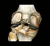
13243021 - Healthy knee, CT scan
14078922 - Flap tear meniscus, illustration
13224583 - Muscles of the back, illustration
13224582 - Muscles of the back, illustration
14078932 - Lateral collateral ligament tear, illustration
14078625 - SLAP lesion of the shoulder, illustration
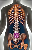
13436088 - Lumber spine, illustration

13435547 - Human muscles, illustration
13435228 - Cervical spine, illustration
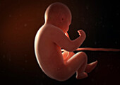
12650244 - Embryo Anatomy, Week 10
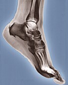
13452940 - Heel pain, X-ray

13435914 - Kidney anatomy, illustration
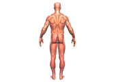
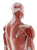
13435761 - Muscular system viewed from behind, illustration
13243022 - Healthy knee, CT scan
14078943 - Meniscus medialis and meniscus lateralis, illustration
13950907 - Corpus callosum, DTI MRI scan
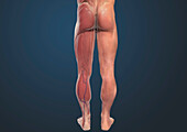
13435968 - Male leg musculature, illustration
13219006 - Lungs affected by Covid-19 atypical pneumonia, CT scans
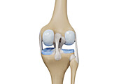
14078925 - Knee, illustration
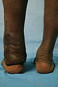
13453022 - Eumycetoma
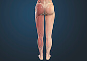
13435961 - Female leg musculature, illustration
13218162 - Lungs affected by Covid-19 atypical pneumonia, 3d CT scan
12918564 - Normal spine, 3d CT scan

12645178 - Testicle anatomy, 1866 illustration
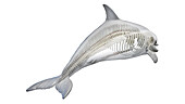
13960998 - Dolphin's skeletal system, illustration
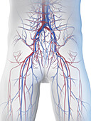
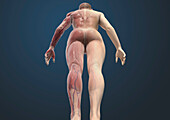
13960799 - Male vascular system, illustration

13960717 - Male skeletal system, illustration
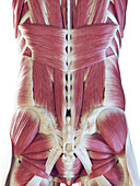
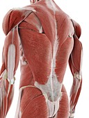
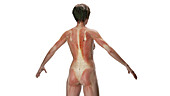
13960679 - Male back muscles, illustration
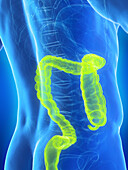
13960673 - Male large intestine, illustration
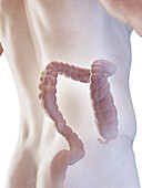
13960663 - Male large intestine, illustration

13960554 - Frog's cardiovascular system, illustration
13960264 - Neck and back anatomy, illustration

13959807 - Abdominal anatomy, illustration
13959439 - Bones of head and upper torso, illustration
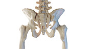
13958958 - Male iliosacral joint, illustration
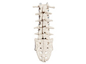
13958919 - Female spine, illustration
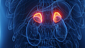
13958816 - Female adrenal glands, illustration
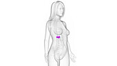
13958815 - Female adrenal glands, illustration
13958647 - Muscular system, illustration
13957472 - Serratus posterior inferior, illustration
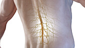
13956895 - Spinal cord, illustration
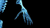
13956837 - Bones of the hand, illustration
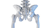
13956769 - Posterior skeletal anatomy of the hip, illustration
13956765 - Posterior skeletal anatomy of the hip, illustration
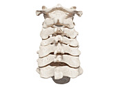
13956729 - Posterior view of the skeletal neck, illustration
page suivante
Postérieur Photos ❘ Science Photo Library