Proton beam therapy for eye tumours
Numéro d’image : 11593384
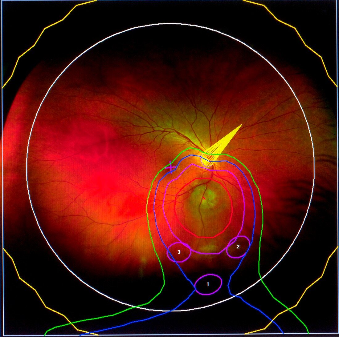
| Retinal camera image,or fundus photograph,overlaid with the data diagram used to prepare the collimator and beam delivery equipment to attack the tumour with great precision. The data seen in the diagram is as follows: red contour line - the base of the tumour,which will receive 100% of the proton dose; purple line - 90% of the dose; blue line - 50% dose; green line - 20% dose. The white line indicates the equator and the numbered areas show the position of the tantalum clips which are placed within the eye to give positional information to align the beam in relation to the tumour. The off-centre yellow feature indicates the position of the macula and fovea,or optic nerve.Photographed at the Clatterbridge Centre for Oncology,Wirral,UK | |
| Licence : | Droits gérés |
| Crédit: | Science Photo Library / King-Holmes, James |
| Taille de l’image : | 4220 px × 4200 px |
| Model Release : | Non requis |
| Property Release : | Non requis |
| Restrictions : | - |
Prix pour cette image À partir de 45 €
Produit vendu
(Calendrier, Carte postale, Carte de vœux, Impression sur textile, Packaging etc)
À partir de 45 €
Usage commercial
(Affichage, Annonce presse, Annonce TV, Carte, Digital - hors rés. sociaux, Digital - rés. sociaux etc)
À partir de 45 €
Éditorial
(Digital, Journal, Livre, Livre pratique, Magazine, Télévision etc)
À partir de 60 €
Usage non-commercial
(Digital - hors rés. sociaux, Digital - rés. sociaux etc)
À partir de 120 €