Pincushion leaf,light micrograph
Numéro d’image : 11562307
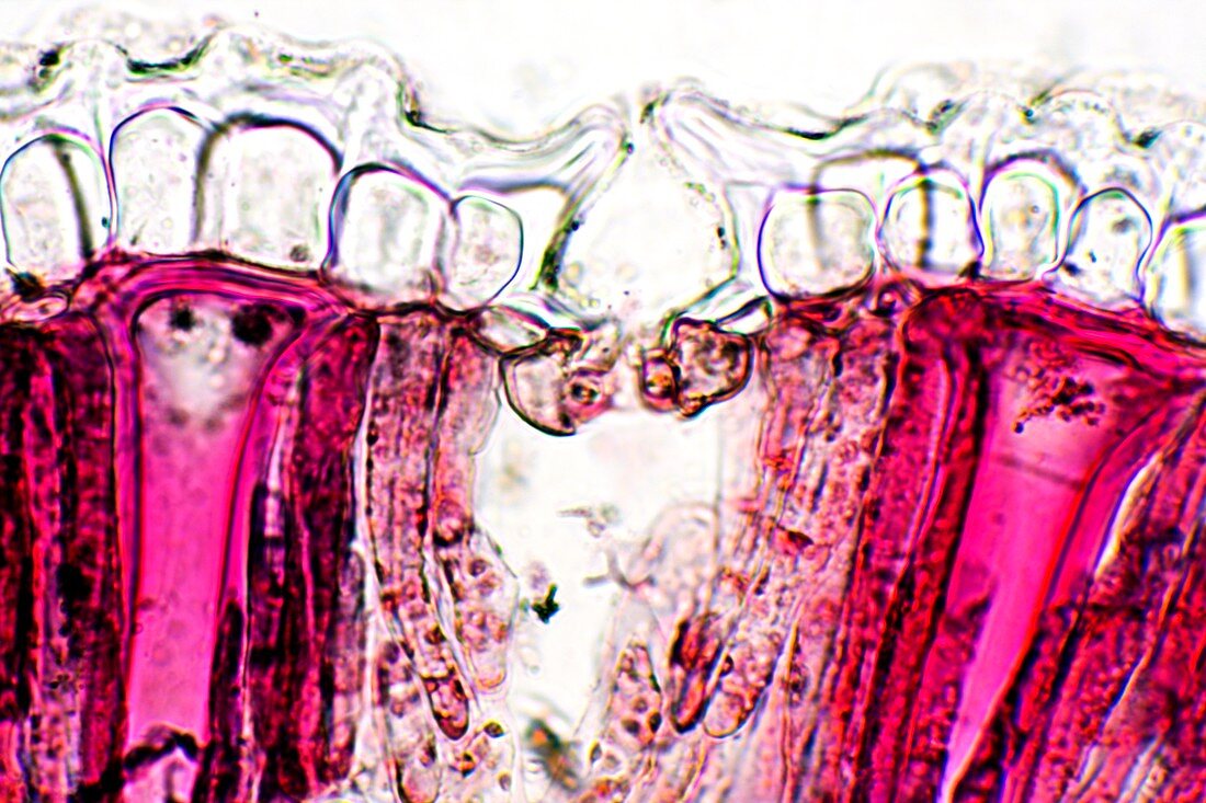
| Pin cushion leaf. Polarised light micrograph of a transverse section through a pinchusion (Hakea laurina) leaf. The leaf of this plant has a number of adaptations that help it to minimise water loss through transpiration. It is covered in a thick cuticle (see-through,top) and its stomata (leaf pores,one seen at centre) are sunken with a 'cup' above them that traps moist air. Also seen are palisade mesophyll cells (red),which contain chloroplasts the site of photosynthesis,and radial sclereid cells (bright pink),thick-walled cells that provide structural support. Magnification: x230 when printed at 10 centimetres wide | |
| Licence : | Droits gérés |
| Crédit: | Science Photo Library / Wheeler, Dr. Keith |
| Taille de l’image : | 5616 px × 3744 px |
| Model Release : | Non requis |
| Property Release : | Non requis |
| Restrictions : | - |
Prix pour cette image À partir de 45 €
Produit vendu
(Calendrier, Carte postale, Carte de vœux, Impression sur textile, Packaging etc)
À partir de 45 €
Usage commercial
(Affichage, Annonce presse, Annonce TV, Carte, Digital - hors rés. sociaux, Digital - rés. sociaux etc)
À partir de 45 €
Éditorial
(Digital, Journal, Livre, Livre pratique, Magazine, Télévision etc)
À partir de 60 €
Usage non-commercial
(Digital - hors rés. sociaux, Digital - rés. sociaux etc)
À partir de 120 €
Mots clés
- adapté à la sécheresse,
- anatomie végétale,
- biologie,
- biologique,
- botanique,
- cellule,
- cellule végétale,
- cellules,
- coupe transversale,
- cuticule,
- divisé,
- feuille,
- flore,
- HAKEA LAURINA,
- micrographe à lumière polarisée,
- microscope à lumière polarisée,
- microscope optique,
- microscopie optique,
- nature,
- palisade mesophyll,
- plante,
- résistant à l'aridité,
- sclérides,
- sclérites,
- section transversale,
- stoma,
- stomates,
- stomie,
- xérophyte