Images
Vidéos
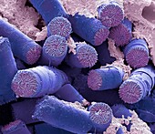
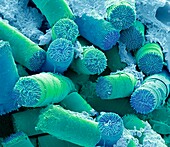
13506576 - Soil diatoms, SEM

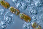
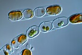
13443452 - Euastrum humerosum desmid, light micrograph
13435588 - Fossil diatom, light micrograph
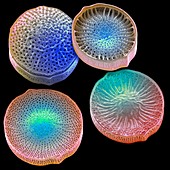
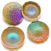
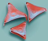
13765677 - Assorted diatoms, light micrograph

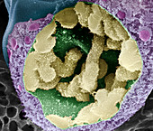
13619590 - Anaphase in onion root tip cell, light micrograph
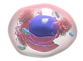
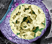
13435225 - Human cell, illustration
12992643 - Diatom, SEM
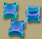
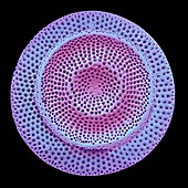
12992641 - Diatoms, SEM
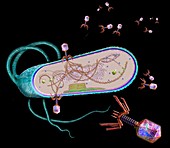
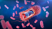
13416736 - Phages infecting a bacterial cell, illustration
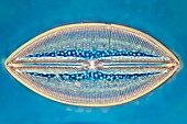
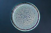
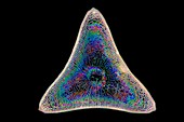
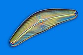
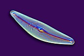
13404287 - Diatom, light micrograph
12992672 - Diatoms, SEM
13403296 - Fossil diatom, light micrograph
12992652 - Diatoms, SEM

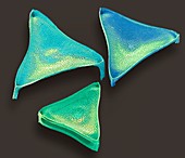
13435598 - Navicula sp. marine diatom, light micrograph
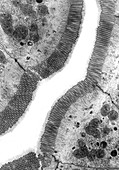
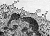
12650137 - Surface Coating of Intestinal Epithelial Cells, EM TEM
12992585 - Diatom, SEM
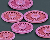
12992580 - Diatoms, SEM
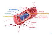
12947764 - Bacterial cell structure, illustration
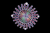
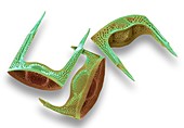
13435632 - Campylodiscus sp. diatoms, light micrograph
12992665 - Diatoms, SEM
12992658 - Diatoms, SEM
12992677 - Diatoms, SEM
12539241 - Trichoderma reesei fungus, TEM
13506575 - Soil diatoms, SEM
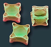
13435624 - Aulacoseira granulata diatoms, light micrograph

13435224 - Human cell, illustration
12992647 - Diatoms, SEM
12649080 - Adipose Tissue, LM
13435625 - Aulacoseira granulata diatoms, light micrograph

13403295 - Fossil diatom, light micrograph
12539240 - Trichoderma reesei fungus, TEM
13506615 - Soil diatoms, SEM
12992632 - Diatom, SEM
12992610 - Diatoms, SEM
12992603 - Diatom, SEM
12650147 - Pinocytosis, TEM
13404304 - Diatom fossil, light micrograph
12992637 - Diatoms, SEM
12992604 - Diatom, SEM

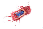
13962958 - Rod-shaped Gram-negative bacterium, illustration
13435587 - Fossil diatom, light micrograph
13272322 - Fossil diatom, light micrograph
13272319 - Diatom, light micrograph
12992593 - Diatoms, SEM
12992581 - Diatom, SEM
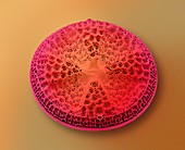
12553827 - Diatom, SEM
12447172 - Diatom, SEM
13403300 - Fossil diatoms, light micrograph
12992588 - Diatom, SEM
12553823 - Diatom, SEM
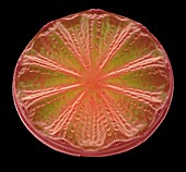
13479965 - Plant cell, light micrograph

12635728 - Plant Cell, Illustration
12553824 - Diatom, SEM
12378132 - Balsa wood (cross section), SEM

12349392 - Wanda K. Farr, US botanist
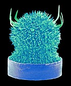
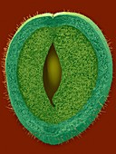
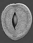
11708517 - Dividing pollen cell,SEM
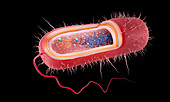
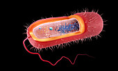
13962953 - Escherichia coli anatomy, illustration

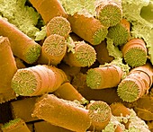
12378188 - Pear sclereids or stone cells (Pyrus sp.), SEM
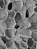
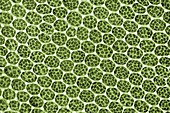
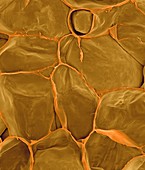
12378177 - Green bean parenchyma cells, SEM
13962955 - Rod-shaped Gram-negative bacterium, illustration
12378182 - Pear fruit parenchyma cells (Pyrus sp.), SEM
12324789 - Diatom, SEM
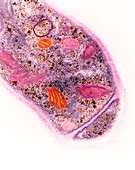
11724854 - Moss leaf,light micrograph
13962954 - Escherichia coli anatomy, illustration
13407576 - Phages infecting a bacterial cell, illustration

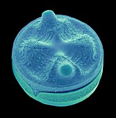
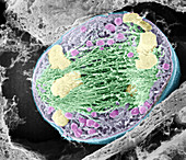
12647990 - Sea Urchin Cell Dividing
12378186 - Pear sclereid or stone cell (Pyrus sp.), SEM
11724857 - Moss leaf,light micrograph
12539242 - Trichoderma reesei fungus, TEM
13495298 - Spinach Leaf Mesophyll, TEM
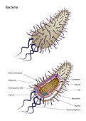
12947763 - Bacterial cell structure, illustration
13962959 - Escherichia coli anatomy, illustration
12650146 - Thrombocyte Cell Structure TEM
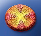
12447206 - Diatom, SEM

12324800 - Diatom, SEM
11708519 - Dividing pollen cell,SEM
13962957 - Rod-shaped Gram-negative bacterium, illustration
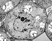
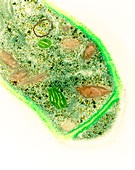
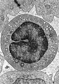
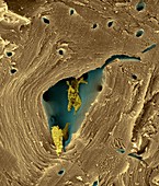
12362450 - Plant parenchyma cell nucleus, TEM
12301693 - Green bean parenchyma cells and seed, SEM
12301692 - Green bean parenchyma cells and seed, SEM
11724860 - Moss leaf,light micrograph
11708513 - Dividing pollen cell,SEM
12378148 - Rosemary leaf cross section (Rosmarinus officinalis), SEM
12301662 - Green bean pod parenchyma cells after steaming., SEM
11724856 - Moss leaf,light micrograph

13962956 - Rod-shaped Gram-negative bacterium, illustration
12301682 - Apple fruit section, skin to parenchyma, SEM
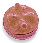
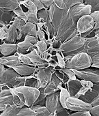
12301587 - Banana fruit cells (Musa sp.), SEM
11727830 - Diatom,light micrograph
12378185 - Green bean parenchyma cells and seed, SEM
12301698 - Pear sclereid or stone cell (Pyrus sp.), SEM
12301674 - Apple parenchyma cells (Malus domestica), SEM
11724851 - Moss leaf,light micrograph
11708530 - Dividing pollen cell,SEM
11708523 - Dividing pollen cell,SEM
12447192 - Diatom, SEM

12301705 - Pear sclereids or stone cells (Pyrus sp.), SEM
12301672 - Pear fruit parenchyma cells (Pyrus sp.), SEM
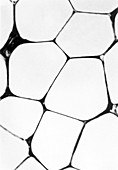
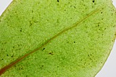
11523597 - LM of cells in a leaf of moss
page suivante
Paroi cellulaire Photos ❘ Science Photo Library