Images
Vidéos

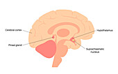
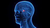
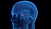
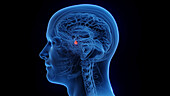
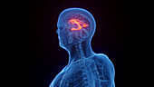
13624678 - Pineal gland, illustration
13624668 - Pineal gland, illustration
13957290 - Tuber cinereum, illustration
13957280 - Optic tract, illustration
13957279 - Optic nerve, illustration
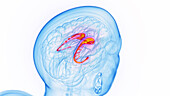
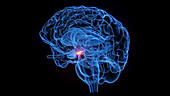
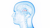
13957220 - Caudate nucleus, illustration
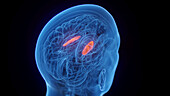
13957218 - Caudate nucleus, illustration
13957266 - Hippocampus, illustration
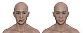
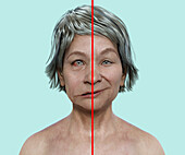
13955490 - Healthy man and man with facial palsy, illustration
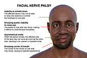
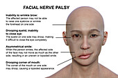
13955488 - Facial palsy, illustration
13624704 - Sleep wake cycle, illustration
13957263 - Fornix crura, illustration
13957259 - Cuneate tubercle and fasciculus, illustration
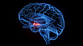
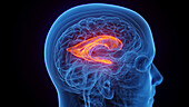
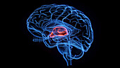
13957252 - Thalamus, illustration
13957232 - Lateral globus pallidus, illustration
13955495 - Facial palsy, illustration
13955497 - Facial palsy, illustration
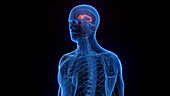
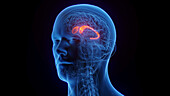
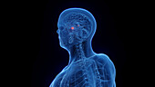
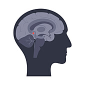
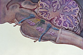
13957226 - Hypothalamus, illustration

12302938 - Elizabeth C. Crosby, American neuroanatomist
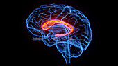
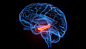
13957254 - Caudate nucleus, illustration
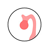
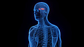
13957244 - Pituitary gland, illustration
13957243 - Pituitary gland, illustration
13957236 - Lateral ventricle, illustration
13957231 - Lateral globus pallidus, illustration
13955499 - Facial palsy, illustration
13957237 - Lateral ventricle, illustration
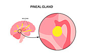
13624680 - Pineal gland, illustration
13957260 - Fornix body, illustration
13957258 - Cerebrum, illustration
13957221 - Hippocampus, illustration
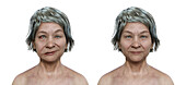
13955492 - Healthy woman and woman with facial palsy, illustration
13957289 - Third ventricle, illustration
13957250 - The putamen, illustration
13957212 - Caudate nucleus, illustration
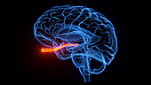
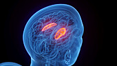
13957209 - Amygdala, illustration
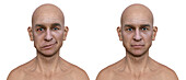
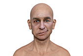
13955491 - Healthy man and man with facial palsy, illustration
13624666 - Pineal gland, illustration

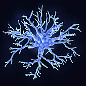
12973666 - Fibrous astrocyte, illustration
13350256 - Protoplasmic astrocyte, illustration
13246599 - Protoplasmic astrocytes, illustration
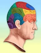
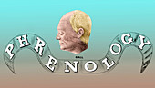
12631072 - Phrenology
13350255 - Protoplasmic astrocyte, illustration
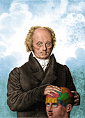
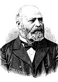
12630952 - Franz Joseph Gall, German Phrenologist
12973665 - Protoplasmic and fibrous astrocytes, illustration
12973668 - Microglial cell, illustration
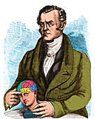
12630953 - Franz Joseph Gall, German Phrenologist
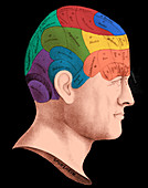
12631071 - Phrenology
12973667 - Protoplasmic astrocyte, illustration
12630954 - Franz Joseph Gall, German Phrenologist
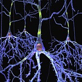
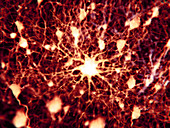
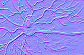
11673125 - Pyramidal cells in the brain,artwork
11659355 - Brain areas,conceptual illustration
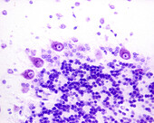
14167540 - Hippocampus, light micrograph

11659519 - Electric ray brain,illustration
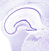
13219518 - Hippocampus, light micrograph
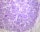
14179811 - Dorsal root ganglion, light micrograph
11665277 - Bernhard von Gudden,German anatomist
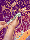
12041787 - Anesthetic,Illustration
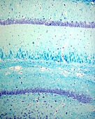
14167583 - Purkinje cells, light micrograph
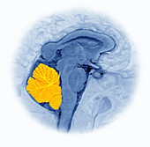
12023233 - Cerebellum
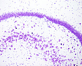
14167580 - Dentate gyrus, light micrograph
12017128 - Brain Stem,Illustration
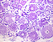
14179812 - Dorsal root ganglion, light micrograph
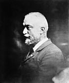
11697058 - August Forel,Swiss psychiatrist
11673126 - Pyramidal cell in the brain,artwork
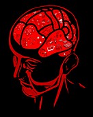
11870535 - Brain map
14179813 - Dorsal root ganglion, light micrograph
11870534 - Brain map
12066416 - Santiago Ramon y Cajal
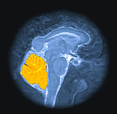
12023234 - Cerebellum
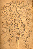
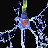
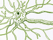
14189517 - Motor neuron structure, illustration
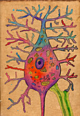
14189519 - Motor neuron structure, illustration
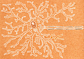
14189526 - Motor neuron structure, illustration
14189521 - Motor neuron structure, illustration
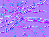
14189525 - Neural network, illustration
14189518 - Motor neuron structure, illustration
14189520 - Motor neuron structure, illustration
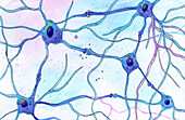
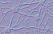
14189524 - Neural network, illustration
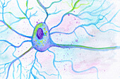
14189522 - Motor neuron structure, illustration
14189523 - Motor neuron structure, illustration
Neuro-anatomie Photos ❘ Science Photo Library