Gastric mucosa, light micrograph
Numéro d’image : 14179802
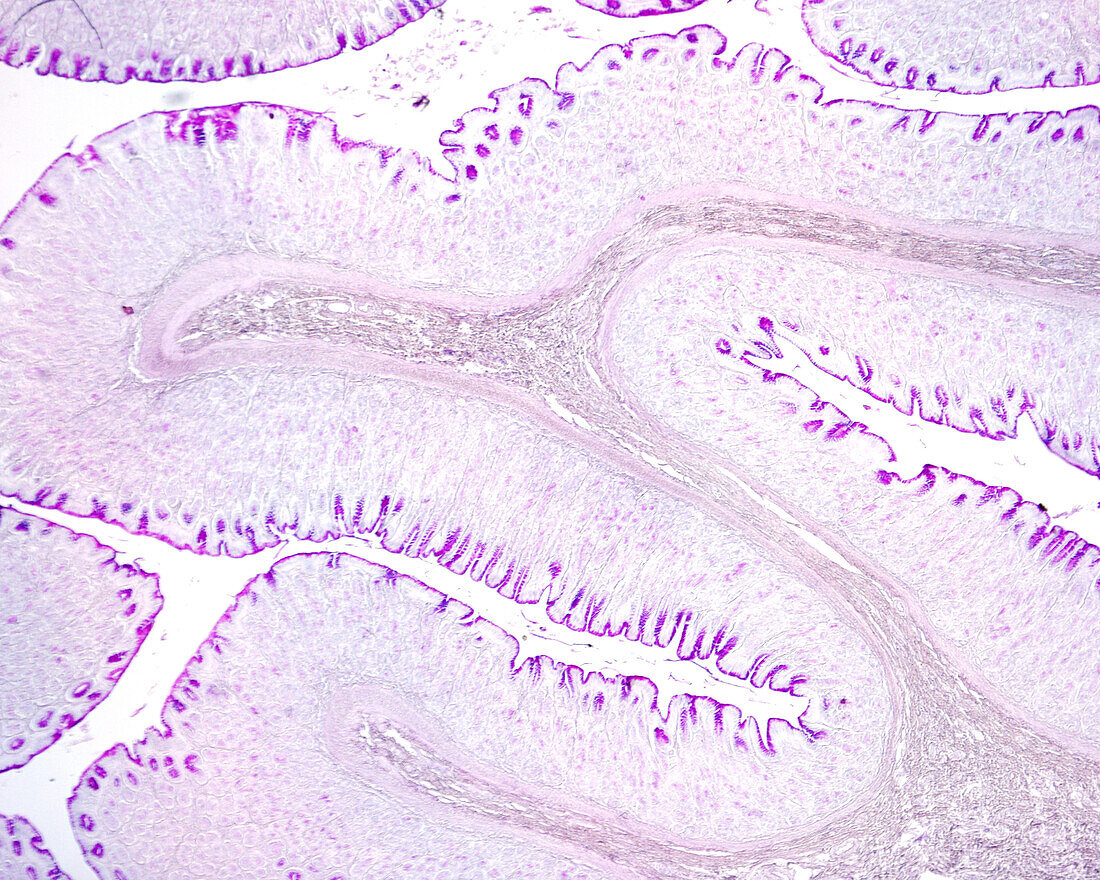
| Light micrograph of the gastric wall stained with the Periodic acid–Schiff (PAS) method. The inner layer is the mucosa that shows many folds. In the axis of these folds is the submucosa. In the mucosa, the mucous surface epithelium and foveolar cells of gastric pits show a great PAS positivity. | |
| Licence : | Droits gérés |
| Crédit: | Science Photo Library / JOSE CALVO |
| Taille de l’image : | 3840 px × 3072 px |
| Model Release : | Non requis |
| Property Release : | Non requis |
| Restrictions : | - |
Prix pour cette image À partir de 45 €
Produit vendu
(Calendrier, Carte postale, Carte de vœux, Impression sur textile, Packaging etc)
À partir de 45 €
Usage commercial
(Affichage, Annonce presse, Annonce TV, Carte, Digital - hors rés. sociaux, Digital - rés. sociaux etc)
À partir de 45 €
Éditorial
(Digital, Journal, Livre, Livre pratique, Magazine, Télévision etc)
À partir de 60 €
Usage non-commercial
(Digital - hors rés. sociaux, Digital - rés. sociaux etc)
À partir de 120 €
Mots clés
- aucun,
- biologie,
- biologique,
- estomac,
- fosse gastrique,
- gastrique,
- histologie,
- histologique,
- micrographie,
- micrographie optique,
- microscope,
- microscope optique,
- microscopie,
- microscopie optique,
- mucosa,
- muqueuse,
- muqueuse gastrique,
- personne,
- sous-muqueux,
- système digestif,
- tissus,
- tractus gastro-intestinal