Human oesophagus, light micrograph
Numéro d’image : 14179796
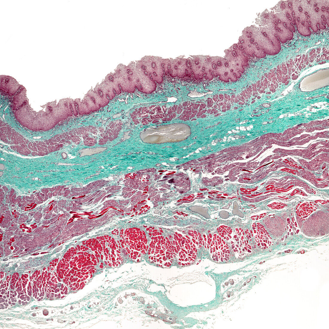
| Light micrograph of a human oesophagus showing, from top, mucosa lined by a stratified squamous epithelium, lamina propria, muscularis mucosae, submucosa, two muscular layers and adventitia. With Masson trichrome stain, the connective (green) and muscular tissue (light purple) differentiate well. | |
| Licence : | Droits gérés |
| Crédit: | Science Photo Library / JOSE CALVO |
| Taille de l’image : | 3840 px × 3835 px |
| Model Release : | Non requis |
| Property Release : | Non requis |
| Restrictions : | - |
Prix pour cette image À partir de 45 €
Produit vendu
(Calendrier, Carte postale, Carte de vœux, Impression sur textile, Packaging etc)
À partir de 45 €
Usage commercial
(Affichage, Annonce presse, Annonce TV, Carte, Digital - hors rés. sociaux, Digital - rés. sociaux etc)
À partir de 45 €
Éditorial
(Digital, Journal, Livre, Livre pratique, Magazine, Télévision etc)
À partir de 60 €
Usage non-commercial
(Digital - hors rés. sociaux, Digital - rés. sociaux etc)
À partir de 120 €
Mots clés
- adventice,
- adventitatia,
- aucun,
- biologie,
- biologique,
- cellule,
- couche musculaire,
- epithelium,
- épithélium,
- esophagus,
- gut,
- histologie,
- histologique,
- lamina propria,
- micrographie,
- micrographie optique,
- microscope,
- microscope optique,
- microscopie,
- microscopie optique,
- mucosa,
- muqueuse,
- muscularis mucosae,
- musculeuse muqeuse,
- œsophage,
- oesophagus,
- personne,
- sous-muqueux,
- tissus,
- tractus digestif,
- tractus gastro-intestinal