Cerebellum, light micrograph
Numéro d’image : 14167570
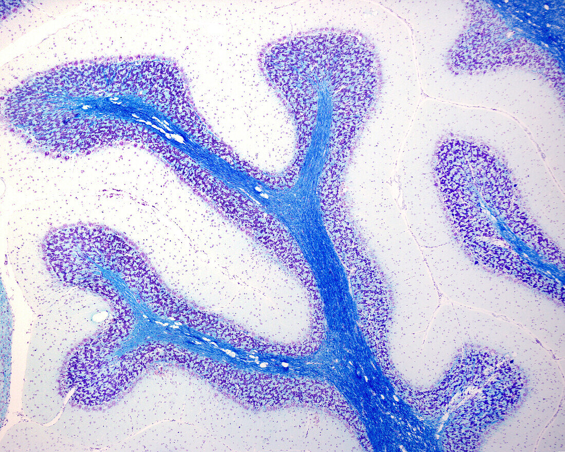
| Light micrograph of a sagittal section of cerebellum stained with Luxol fast blue and cresyl violet. Each cerebellar folium shows the molecular and granular layers and the central axis of white matter with blue stained myelinated fibres. | |
| Licence : | Droits gérés |
| Crédit: | Science Photo Library / JOSE CALVO |
| Taille de l’image : | 3840 px × 3072 px |
| Model Release : | Non requis |
| Property Release : | Non requis |
| Restrictions : | - |
Prix pour cette image À partir de 45 €
Produit vendu
(Calendrier, Carte postale, Carte de vœux, Impression sur textile, Packaging etc)
À partir de 45 €
Usage commercial
(Affichage, Annonce presse, Annonce TV, Carte, Digital - hors rés. sociaux, Digital - rés. sociaux etc)
À partir de 45 €
Éditorial
(Digital, Journal, Livre, Livre pratique, Magazine, Télévision etc)
À partir de 60 €
Usage non-commercial
(Digital - hors rés. sociaux, Digital - rés. sociaux etc)
À partir de 120 €
Mots clés
- aucun,
- biologie,
- biologique,
- cerebellum,
- cervelet,
- couche de molécules,
- couche granulaire,
- couche moléculaire,
- en bonne santé,
- fibre,
- histologie,
- histologique,
- matière grise,
- micrographie,
- micrographie optique,
- microscope,
- microscope optique,
- microscopie,
- microscopie optique,
- microscopique,
- molécule,
- myélinisé,
- nerf,
- nerveux,
- neurohistologie,
- neurologie,
- neurologique,
- normal,
- personne,
- S.N.C.,
- sain,
- SNC,
- système nerveux central