Epiphyseal growth plate, light micrograph
Numéro d’image : 14167556
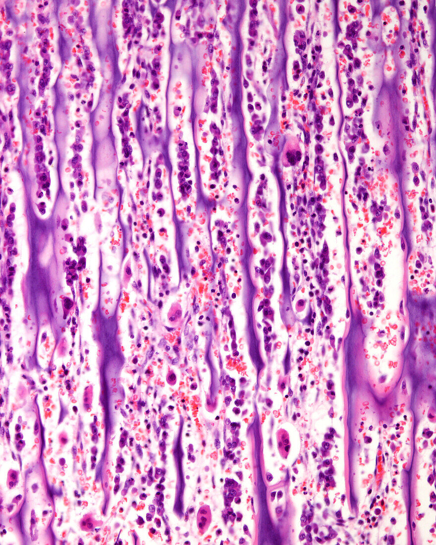
| Endochondral ossification, light micrograph. Zone of ossification of a growth plate. Mixed trabeculae with a centre of calcified hyaline cartilage (blue) are surrounded by mineralised bone tissue (pink). A layer of osteoblasts can be seen surrounding the trabeculae. | |
| Licence : | Droits gérés |
| Crédit: | Science Photo Library / JOSE CALVO |
| Taille de l’image : | 3072 px × 3840 px |
| Model Release : | Non requis |
| Property Release : | Non requis |
| Restrictions : | - |
Prix pour cette image À partir de 45 €
Produit vendu
(Calendrier, Carte postale, Carte de vœux, Impression sur textile, Packaging etc)
À partir de 45 €
Usage commercial
(Affichage, Annonce presse, Annonce TV, Carte, Digital - hors rés. sociaux, Digital - rés. sociaux etc)
À partir de 45 €
Éditorial
(Digital, Journal, Livre, Livre pratique, Magazine, Télévision etc)
À partir de 60 €
Usage non-commercial
(Digital - hors rés. sociaux, Digital - rés. sociaux etc)
À partir de 120 €
Mots clés
- aucun,
- biologie,
- biologique,
- cartilage,
- cartilage de conjugaison,
- cellule,
- chondrocytes,
- en bonne santé,
- épiphyse,
- histologie,
- histologique,
- métaphyse,
- micrographie,
- micrographie optique,
- microscope,
- microscope optique,
- microscopie,
- microscopie optique,
- moelle osseuse,
- normal,
- os,
- ossification,
- personne,
- plaque de croissane,
- sain,
- sclérose,
- tissus