Normal coronal and cross-sections of brain, illustration
Numéro d’image : 14167136
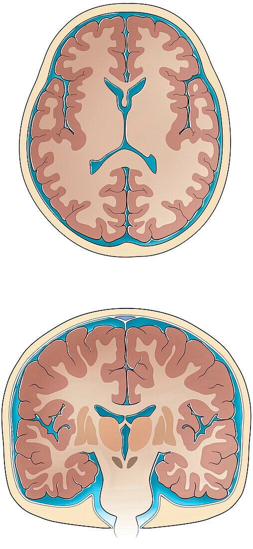
| Normal coronal and cross-sections of brain, illustration. Slices through the normal brain and skull showing ventricles, white matter, grey matter, basal ganglia, thalamus sulci and gyri, cerebrospinal fluid and skull. | |
| Licence : | Droits gérés |
| Crédit: | Science Photo Library / Sue Seif |
| Taille de l’image : | 2841 px × 6150 px |
| Model Release : | Non requis |
| Property Release : | Non requis |
| Restrictions : | - |
Prix pour cette image À partir de 45 €
Produit vendu
(Calendrier, Carte postale, Carte de vœux, Impression sur textile, Packaging etc)
À partir de 45 €
Usage commercial
(Affichage, Annonce presse, Annonce TV, Carte, Digital - hors rés. sociaux, Digital - rés. sociaux etc)
À partir de 45 €
Éditorial
(Digital, Journal, Livre, Livre pratique, Magazine, Télévision etc)
À partir de 60 €
Usage non-commercial
(Digital - hors rés. sociaux, Digital - rés. sociaux etc)
À partir de 120 €
Mots clés
- anatomie,
- anatomique,
- aucun,
- basal,
- basale,
- base,
- blanc,
- cérébrospinal,
- cerveau,
- circonvolution cérébrale,
- coronaire,
- coronal,
- coronale,
- coupe transversale,
- crâne,
- dessin,
- dessins,
- fluide,
- ganglions,
- gris,
- illustration,
- illustrations,
- matière,
- normal,
- oeuvre,
- personne,
- sections transversales,
- sillons,
- sulci,
- thalamus,
- ventricules