Organ of Corti, light micrograph
Numéro d’image : 14114319
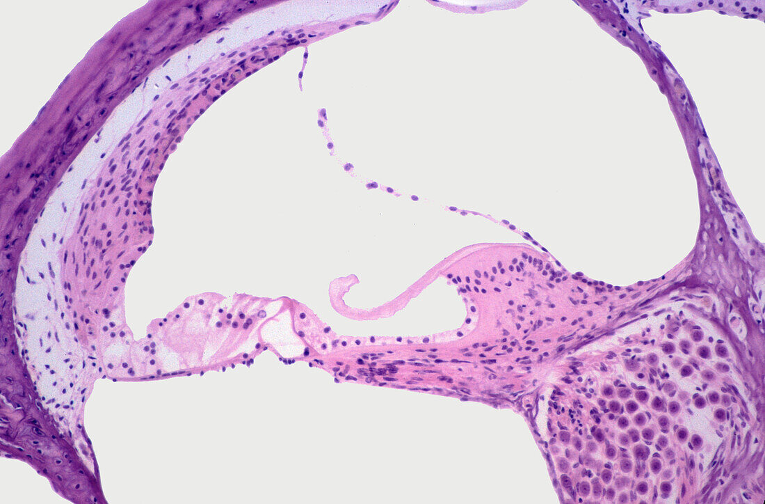
| Light micrograph of a cross-section of the cochlea of the inner ear showing from top to bottom: the vestibular, cochlear and tympanic ducts, or scala tympani. The cochlear duct shows, from left to right: the spiral ganglion, limbus spiralis with tectorial membrane, organ of Corti with tunnel of Corti and hair cells, and the stria vascularis. | |
| Licence : | Droits gérés |
| Crédit: | Science Photo Library / JOSE CALVO |
| Taille de l’image : | 4769 px × 3139 px |
| Model Release : | Non requis |
| Property Release : | Non requis |
| Restrictions : | - |
Prix pour cette image À partir de 45 €
Produit vendu
(Calendrier, Carte postale, Carte de vœux, Impression sur textile, Packaging etc)
À partir de 45 €
Usage commercial
(Affichage, Annonce presse, Annonce TV, Carte, Digital - hors rés. sociaux, Digital - rés. sociaux etc)
À partir de 45 €
Éditorial
(Digital, Journal, Livre, Livre pratique, Magazine, Télévision etc)
À partir de 60 €
Usage non-commercial
(Digital - hors rés. sociaux, Digital - rés. sociaux etc)
À partir de 120 €
Mots clés
- anatomie,
- anatomique,
- arrière plan blanc,
- arrière-plan blanc,
- aucun,
- audio,
- auditif,
- audition,
- biologie,
- biologique,
- cochlea,
- cochlée,
- en bonne santé,
- fond blanc,
- histologie,
- histologique,
- membrane tectorielle,
- micrographie,
- microscope,
- microscope optique,
- microscopie,
- microscopie optique,
- normal,
- oral,
- oreille,
- oreille interne,
- organe sensoriel,
- otologie,
- personne,
- sain,
- sonore