Pilar cyst, light micrograph
Numéro d’image : 14113492
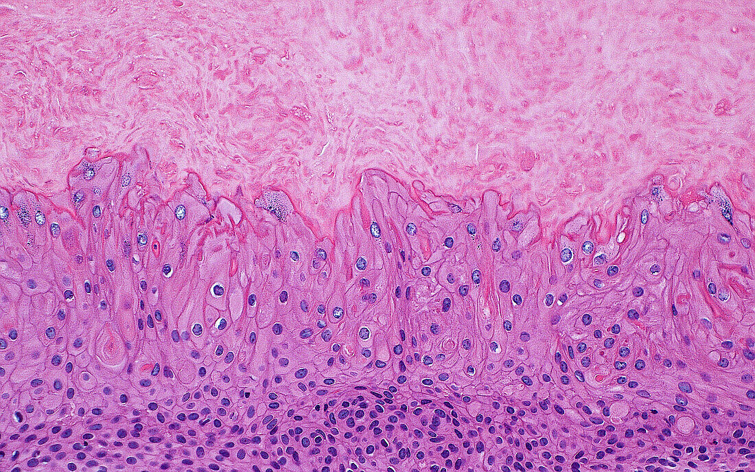
| Light micrograph of a pilar cyst, which is a benign skin occurring in skin. The cyst lining cells are squamous cells (bottom half of image) and the cyst contents are dense keratin material (light pink at top half of image). The squamous cells contain nuclei (round purple dots), whereas the keratin is void of nuclei. Haematoxylin and eosin stained tissue section. Magnification: 200x when printed at 10cm. | |
| Licence : | Droits gérés |
| Crédit: | Science Photo Library / ZIAD M. EL-ZAATARI |
| Taille de l’image : | 4096 px × 2560 px |
| Model Release : | Non requis |
| Property Release : | Non requis |
| Restrictions : | - |
Prix pour cette image À partir de 45 €
Produit vendu
(Calendrier, Carte postale, Carte de vœux, Impression sur textile, Packaging etc)
À partir de 45 €
Usage commercial
(Affichage, Annonce presse, Annonce TV, Carte, Digital - hors rés. sociaux, Digital - rés. sociaux etc)
À partir de 45 €
Éditorial
(Digital, Journal, Livre, Livre pratique, Magazine, Télévision etc)
À partir de 60 €
Usage non-commercial
(Digital - hors rés. sociaux, Digital - rés. sociaux etc)
À partir de 120 €
Mots clés
- anatomie,
- anatomie pathologique,
- anormal,
- aucun,
- cellules,
- corps humain,
- dermatologie,
- désordre,
- état,
- glisser,
- histopathologie,
- histopathologique,
- maladie,
- malsain,
- médecine,
- médical,
- médicale,
- micrographie optique,
- microscope,
- microscope optique,
- microscopie,
- microscopie optique,
- pathologie,
- pathologie anatomique,
- pathologique,
- peau,
- personne,
- tissus,
- trouble,
- tumeur nénigne