Liver cirrhosis, light micrograph
Numéro d’image : 14113485
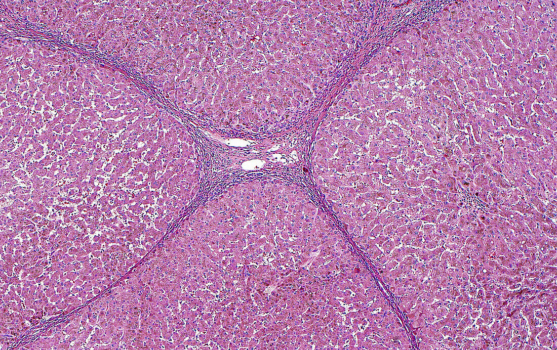
| Light micrograph of cirrhosis. In cirrhosis, the end stage of chronic liver damage, the liver cells are arranged in nodules surrounded by fibrous septa (bands). In this image, the intersection (centre of image) of four nodules and their surrounding septa are seen. Haematoxylin and eosin stained tissue section. Magnification: 40x when printed at 10cm. | |
| Licence : | Droits gérés |
| Crédit: | Science Photo Library / ZIAD M. EL-ZAATARI |
| Taille de l’image : | 4083 px × 2570 px |
| Model Release : | Non requis |
| Property Release : | Non requis |
| Restrictions : | - |
Prix pour cette image À partir de 45 €
Produit vendu
(Calendrier, Carte postale, Carte de vœux, Impression sur textile, Packaging etc)
À partir de 45 €
Usage commercial
(Affichage, Annonce presse, Annonce TV, Carte, Digital - hors rés. sociaux, Digital - rés. sociaux etc)
À partir de 45 €
Éditorial
(Digital, Journal, Livre, Livre pratique, Magazine, Télévision etc)
À partir de 60 €
Usage non-commercial
(Digital - hors rés. sociaux, Digital - rés. sociaux etc)
À partir de 120 €
Mots clés
- anatomie,
- anatomie pathologique,
- anormal,
- aucun,
- cellules,
- cellules du foie,
- cirrhose,
- cirrhosis,
- corps humain,
- désordre,
- état,
- foie,
- glisser,
- hépatocyte,
- hépatologie,
- histopathologie,
- histopathologique,
- maladie,
- maladie du foie,
- malsain,
- médecine,
- médical,
- médicale,
- micrographie optique,
- microscope,
- microscope optique,
- microscopie,
- microscopie optique,
- pathologie,
- pathologie anatomique,
- pathologique,
- personne,
- tissus,
- trouble