Breast cyst secretions, light micrograph
Numéro d’image : 14113444
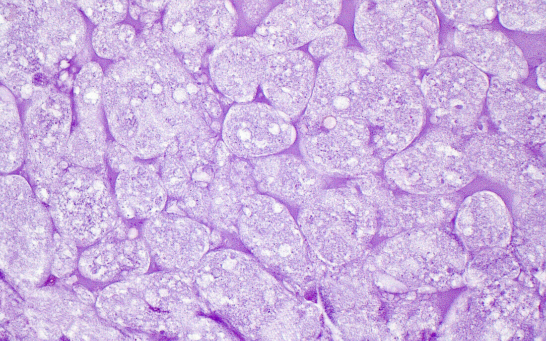
| Light micrograph of secretions within a breast microcyst. Only the secretions (which here resemble puffs or clouds) that were inside the cyst are seen in this image, and not the cyst wall or lining cells. Haematoxylin and eosin stained tissue section. Magnification: 400x when printed at 10cm. | |
| Licence : | Droits gérés |
| Crédit: | Science Photo Library / ZIAD M. EL-ZAATARI |
| Taille de l’image : | 4096 px × 2560 px |
| Model Release : | Non requis |
| Property Release : | Non requis |
| Restrictions : | - |
Prix pour cette image À partir de 45 €
Produit vendu
(Calendrier, Carte postale, Carte de vœux, Impression sur textile, Packaging etc)
À partir de 45 €
Usage commercial
(Affichage, Annonce presse, Annonce TV, Carte, Digital - hors rés. sociaux, Digital - rés. sociaux etc)
À partir de 45 €
Éditorial
(Digital, Journal, Livre, Livre pratique, Magazine, Télévision etc)
À partir de 60 €
Usage non-commercial
(Digital - hors rés. sociaux, Digital - rés. sociaux etc)
À partir de 120 €
Mots clés
- anatomie,
- anatomie pathologique,
- anormal,
- aucun,
- bénin,
- cellules,
- corps humain,
- désordre,
- état,
- glisser,
- histopathologie,
- histopathologique,
- kyste,
- maladie,
- malsain,
- médecine,
- médical,
- médicale,
- micrographie optique,
- microscope,
- microscope optique,
- microscopie,
- microscopie optique,
- pathologie,
- pathologie anatomique,
- pathologique,
- personne,
- poitrine,
- sein,
- tissus,
- trouble