Vas deferens smooth muscle, light micrograph
Numéro d’image : 14113439
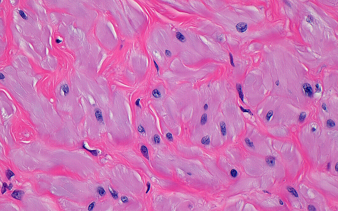
| Light micrograph of smooth muscle cells in the wall of a vas deferens. Groups of smooth muscle cells (pale pink) are surrounded by fibrous tissue (darker pink). The nuclei of the smooth muscle cells are the blue-purple dots. Haematoxylin and eosin stained tissue section. Magnification: 400x when printed at 10cm. | |
| Licence : | Droits gérés |
| Crédit: | Science Photo Library / ZIAD M. EL-ZAATARI |
| Taille de l’image : | 4096 px × 2560 px |
| Model Release : | Non requis |
| Property Release : | Non requis |
| Restrictions : | - |
Prix pour cette image À partir de 45 €
Produit vendu
(Calendrier, Carte postale, Carte de vœux, Impression sur textile, Packaging etc)
À partir de 45 €
Usage commercial
(Affichage, Annonce presse, Annonce TV, Carte, Digital - hors rés. sociaux, Digital - rés. sociaux etc)
À partir de 45 €
Éditorial
(Digital, Journal, Livre, Livre pratique, Magazine, Télévision etc)
À partir de 60 €
Usage non-commercial
(Digital - hors rés. sociaux, Digital - rés. sociaux etc)
À partir de 120 €
Mots clés
- anatomie,
- anatomique,
- aucun,
- biologie,
- biologique,
- canal déférent,
- cellules,
- corps humain,
- en bonne santé,
- fécondité,
- génito-urinaire,
- glisser,
- histologie,
- histologique,
- micrographie optique,
- microscope,
- microscope optique,
- microscopie,
- microscopie optique,
- muscle lisse,
- normal,
- personne,
- reproduction,
- sain,
- système reproducteur masculin,
- tissus,
- urologie,
- vasectomie