Small intestine villi, light micrograph
Numéro d’image : 13952295
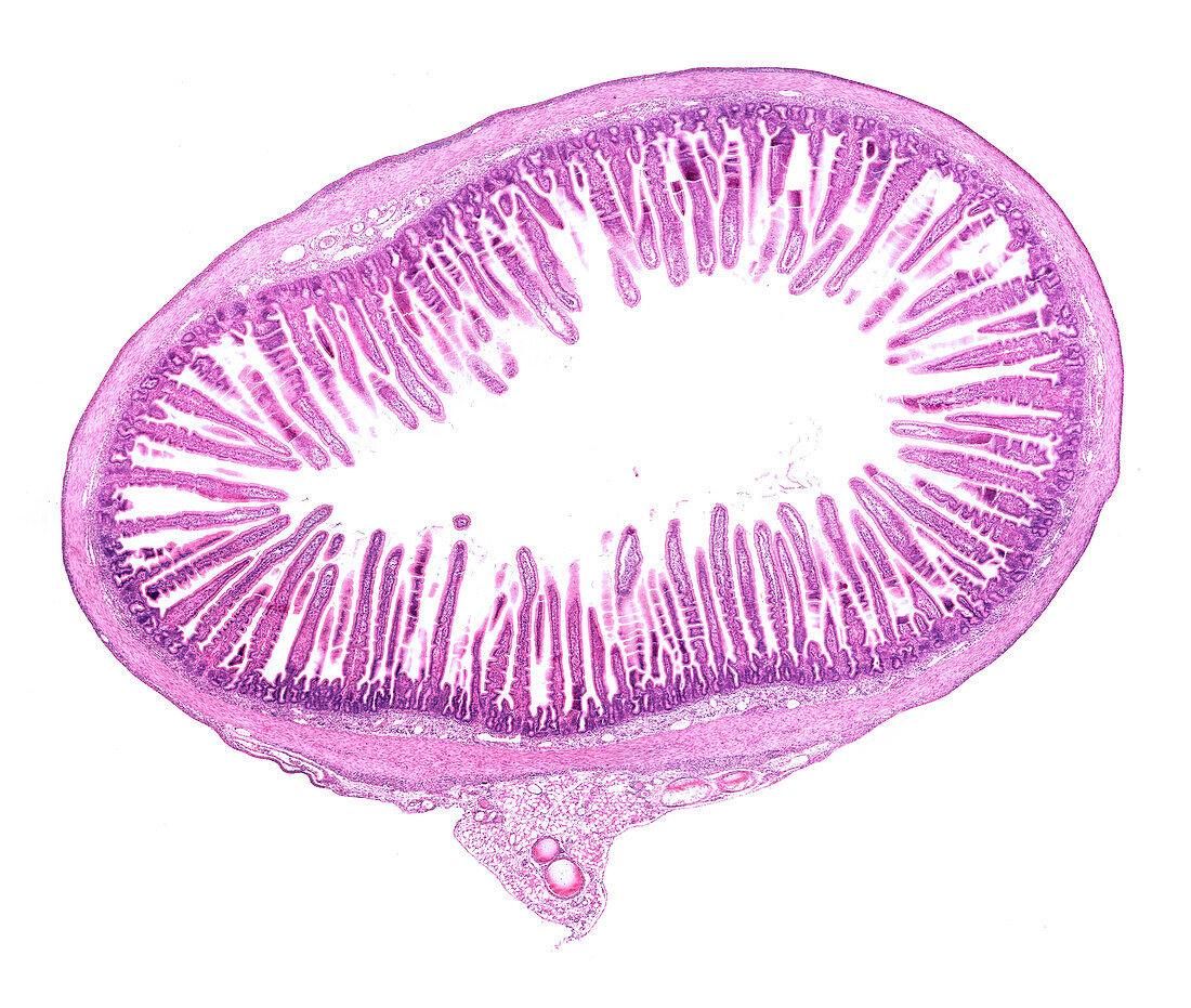
| Light micrograph of a cross-sectioned rat small intestine. The mucosa layer shows abundant villi, finger-like projections that extend into the lumen. Outside the mucosa, the submucosa and the muscular layer can be seen. The mesentery, with blood vessels (round), is at bottom centre. | |
| Licence : | Droits gérés |
| Crédit: | Science Photo Library / JOSE CALVO |
| Taille de l’image : | 3940 px × 3333 px |
| Model Release : | Non requis |
| Property Release : | Non requis |
| Restrictions : | - |
Prix pour cette image À partir de 45 €
Produit vendu
(Calendrier, Carte postale, Carte de vœux, Impression sur textile, Packaging etc)
À partir de 45 €
Usage commercial
(Affichage, Annonce presse, Annonce TV, Carte, Digital - hors rés. sociaux, Digital - rés. sociaux etc)
À partir de 45 €
Éditorial
(Digital, Journal, Livre, Livre pratique, Magazine, Télévision etc)
À partir de 60 €
Usage non-commercial
(Digital - hors rés. sociaux, Digital - rés. sociaux etc)
À partir de 120 €
Mots clés
- aucun,
- biologie,
- biologique,
- en bonne santé,
- epithelium,
- épithélium,
- gut,
- histologie,
- histologique,
- intestin,
- intestin grêle,
- micrographie optique,
- microscope,
- microscope optique,
- microscopie,
- microscopie optique,
- normal,
- personne,
- sain,
- tissus,
- tractus digestif,
- tractus gastro-intestinal,
- villosités