Solitary fibrous tumour vessels, light micrograph
Numéro d’image : 13950523
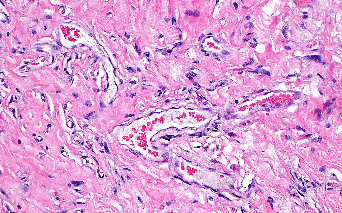
| Light micrograph of a solitary fibrous tumour, a type of growth that can occur in the soft tissues of the body. The tumour cells have spindled nuclei (dark purple) and are present within variable amounts of collagen substance (pink). Another characteristic of solitary fibrous tumours is the presence of dilated (widened) and branching vessels, a few of which are seen in this image. The vessels have lumens (inner white spaces) containing several red blood cells (small bright red dots). Haematoxylin and eosin stained tissue section. Magnification: x200 when printed at 10cm wide. | |
| Licence : | Droits gérés |
| Crédit: | Science Photo Library / ZIAD M. EL-ZAATARI |
| Taille de l’image : | 5000 px × 3124 px |
| Model Release : | Non requis |
| Property Release : | Non requis |
| Restrictions : | - |
Prix pour cette image À partir de 45 €
Produit vendu
(Calendrier, Carte postale, Carte de vœux, Impression sur textile, Packaging etc)
À partir de 45 €
Usage commercial
(Affichage, Annonce presse, Annonce TV, Carte, Digital - hors rés. sociaux, Digital - rés. sociaux etc)
À partir de 45 €
Éditorial
(Digital, Journal, Livre, Livre pratique, Magazine, Télévision etc)
À partir de 60 €
Usage non-commercial
(Digital - hors rés. sociaux, Digital - rés. sociaux etc)
À partir de 120 €
Mots clés
- anatomie,
- anatomie pathologique,
- aucun,
- biologie,
- cellules,
- corps humain,
- glisser,
- hématoxyline,
- histologie,
- histologique,
- histopathologie,
- médecine,
- médical,
- médicale,
- micrographie optique,
- microscope,
- microscope optique,
- microscopie,
- microscopie optique,
- néoplasme,
- pathologie,
- pathologie anatomique,
- pathologique,
- personne,
- tissu mou,
- tissus,
- tumeur,
- vaisseaux