Sage leaf, SEM
Numéro d’image : 13756267
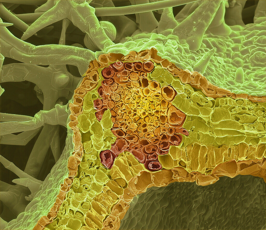
| Sage leaf. Coloured scanning electron micrograph (SEM) of a freeze-fracture through the mid rib of a sage leaf (Salvia officinalis). The large central vascular bundle has contains xylem surrounded by phloem. Smaller vascular bundles are also visible. Below the upper epidermis and surrounding the vascular bundles is a layer of palisade parenchyma cells containing chloroplasts. Beneath this layer is the spongy mesophyll which has large intracellular spaces for gaseous exchange. At the bottom is the lower epidermis, a single layer of closely packed cells where the stomata control gas exchange within the spongy mesophyll. Sage leaves are used in cooking and herbal medicine. Magnification: x35 when printed 10 cm wide. | |
| Licence : | Droits gérés |
| Crédit: | Science Photo Library / Gschmeissner, Steve |
| Taille de l’image : | 4572 px × 3956 px |
| Model Release : | Non requis |
| Restrictions : | - |
Prix pour cette image À partir de 45 €
Produit vendu
(Calendrier, Carte postale, Carte de vœux, Impression sur textile, Packaging etc)
À partir de 45 €
Usage commercial
(Affichage, Annonce presse, Annonce TV, Carte, Digital - hors rés. sociaux, Digital - rés. sociaux etc)
À partir de 45 €
Éditorial
(Digital, Journal, Livre, Livre pratique, Magazine, Télévision etc)
À partir de 60 €
Usage non-commercial
(Digital - hors rés. sociaux, Digital - rés. sociaux etc)
À partir de 120 €