Joint synovial membrane, light micrograph
Numéro d’image : 13756180
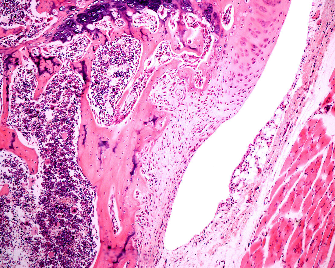
| Light micrograph of a synovial membrane (innermost part of the joint capsule). It delimits the joint cavity of the diarthrosis. A band of synovial cells is concentrated in the vicinity of the lumen of the joint cavity, adopting an appearance reminiscent of a lining epithelium, however, these synovial cells are connective tissue cells that form a discontinuous lining. Deeper is a space occupied by loose connective tissue and developing bone (left) or muscle (right). At top, the transition between synovial membrane and articular cartilage can be seen. | |
| Licence : | Droits gérés |
| Crédit: | Science Photo Library / JOSE CALVO |
| Taille de l’image : | 3840 px × 3072 px |
| Model Release : | Non requis |
| Property Release : | Non requis |
| Restrictions : | - |
Prix pour cette image À partir de 45 €
Produit vendu
(Calendrier, Carte postale, Carte de vœux, Impression sur textile, Packaging etc)
À partir de 45 €
Usage commercial
(Affichage, Annonce presse, Annonce TV, Carte, Digital - hors rés. sociaux, Digital - rés. sociaux etc)
À partir de 45 €
Éditorial
(Digital, Journal, Livre, Livre pratique, Magazine, Télévision etc)
À partir de 60 €
Usage non-commercial
(Digital - hors rés. sociaux, Digital - rés. sociaux etc)
À partir de 120 €