Neurosecretion in posterior pituitary gland, TEM
Numéro d’image : 13755942
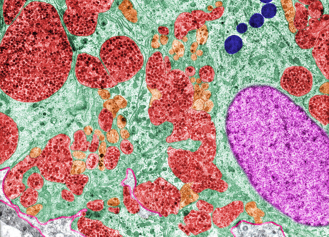
| Coloured transmission electron micrograph (TEM) of the posterior pituitary gland showing axons (red) of hypothalamic neurons (nerve cells) full of neurosecretory granules containing oxytocin and vasopressin. Among them there are pituicytes (light green), a specialised glial cell, that may show lipid droplets (dark blue), a feature typical of pituicytes. The basement membrane is pink. | |
| Licence : | Droits gérés |
| Crédit: | Science Photo Library / JOSE CALVO |
| Taille de l’image : | 4251 px × 3072 px |
| Model Release : | Non requis |
| Property Release : | Non requis |
| Restrictions : | - |
Prix pour cette image À partir de 45 €
Produit vendu
(Calendrier, Carte postale, Carte de vœux, Impression sur textile, Packaging etc)
À partir de 45 €
Usage commercial
(Affichage, Annonce presse, Annonce TV, Carte, Digital - hors rés. sociaux, Digital - rés. sociaux etc)
À partir de 45 €
Éditorial
(Digital, Journal, Livre, Livre pratique, Magazine, Télévision etc)
À partir de 60 €
Usage non-commercial
(Digital - hors rés. sociaux, Digital - rés. sociaux etc)
À partir de 120 €
Mots clés
- aucun,
- coloré,
- colorié,
- colorisé,
- endocrine,
- endocrinologie,
- glande,
- glandulaire,
- histologie,
- histologique,
- hormone,
- humain,
- hypophyse,
- hypophyse postérieure,
- M.E.T.,
- MET,
- micrographie électronique à transmission,
- microscope électronique,
- microscope électronique à transmission,
- neurohypophyse,
- personne,
- posthypophyse,
- ultrastructure