Human chromosomes, light micrograph
Numéro d’image : 13755837
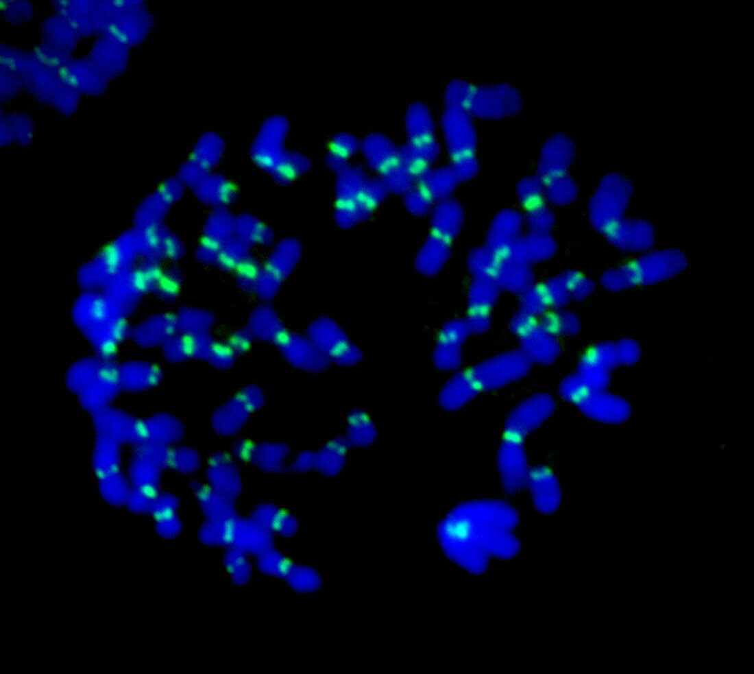
| Fluorescent light micrograph of human chromosomes. Fluorescent markers have been used to highlight DNA (deoxyribonucleic acid, blue) and kinetochores (green). During mitosis (nuclear division), the formation of two daughter nuclei from one parent nucleus, the spindle apparatus that separates' sister chromatids attaches to the chromosomes via the kinetochores. Two identical chromatids make up one chromosome, so each cell retains a copy of the parent cell's genetic information. | |
| Licence : | Droits gérés |
| Crédit: | Science Photo Library / DR. JUAN F. GIMENEZ-ABIAN |
| Taille de l’image : | 3140 px × 2806 px |
| Model Release : | Non requis |
| Property Release : | Non requis |
| Restrictions : | - |
Prix pour cette image À partir de 45 €
Produit vendu
(Calendrier, Carte postale, Carte de vœux, Impression sur textile, Packaging etc)
À partir de 45 €
Usage commercial
(Affichage, Annonce presse, Annonce TV, Carte, Digital - hors rés. sociaux, Digital - rés. sociaux etc)
À partir de 45 €
Éditorial
(Digital, Journal, Livre, Livre pratique, Magazine, Télévision etc)
À partir de 60 €
Usage non-commercial
(Digital - hors rés. sociaux, Digital - rés. sociaux etc)
À partir de 120 €
Mots clés
- A.D.N.,
- acide désoxyribonucléique,
- ADN,
- arrière plan noir,
- arrière-plan noir,
- aucun,
- biologie,
- biologie cellulaire,
- biologique,
- cellule,
- cellules,
- centrosome,
- chromatide,
- chromatides,
- chromosome,
- chromosomes,
- condensé,
- cycle cellulaire,
- cytologie,
- cytologique,
- fluorescence,
- fluorescent,
- fond noir,
- génétique,
- humain,
- kinétochore,
- micrographie optique,
- microscope,
- microscope optique,
- microscopie,
- microscopie optique,
- personne