Harlequin chromosomes, light micrograph
Numéro d’image : 13754754
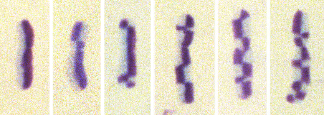
| Light micrograph of six chromosomes from an onion (Allium cepa) cell showing different degrees of harlequin banding. Chromosomes are a packaged form of a cell's genetic material DNA (deoxyribonucleic acid). The DNA condenses into chromosomes during cell replication. Each chromosome consists of two identical strands (chromatids), aligned parallel to each other and joined at an area called the centromere. The harlequin staining technique shows which chromatid was formed first (dark purple) and which second (light purple). Occasionally sister chromatids may exchange segments, particularly in the presence of genotoxic substances, resulting in the banded patterns seen in this micrograph. | |
| Licence : | Droits gérés |
| Crédit: | Science Photo Library / DR. JUAN F. GIMENEZ-ABIAN |
| Taille de l’image : | 4956 px × 1767 px |
| Model Release : | Non requis |
| Property Release : | Non requis |
| Restrictions : | - |
Prix pour cette image À partir de 45 €
Produit vendu
(Calendrier, Carte postale, Carte de vœux, Impression sur textile, Packaging etc)
À partir de 45 €
Usage commercial
(Affichage, Annonce presse, Annonce TV, Carte, Digital - hors rés. sociaux, Digital - rés. sociaux etc)
À partir de 45 €
Éditorial
(Digital, Journal, Livre, Livre pratique, Magazine, Télévision etc)
À partir de 60 €
Usage non-commercial
(Digital - hors rés. sociaux, Digital - rés. sociaux etc)
À partir de 120 €
Mots clés
- à bande,
- A.D.N.,
- acide désoxyribonucléique,
- ADN,
- ALLIUM CEPA,
- aucun,
- bander,
- bandes,
- biologie,
- biologique,
- calicot de feu,
- cercler,
- chromatide,
- chromosome,
- chromosomes,
- chromosomique,
- colorer,
- en bande,
- formation,
- génétique,
- harlequin,
- micrographie optique,
- microscope optique,
- microscope photonique,
- microscopie optique,
- microscopie photonique,
- oignon,
- personne,
- rubané,
- tache,
- technique,
- virus du chou arlequin