Simple columnar epithelium, light micrograph
Numéro d’image : 13742445
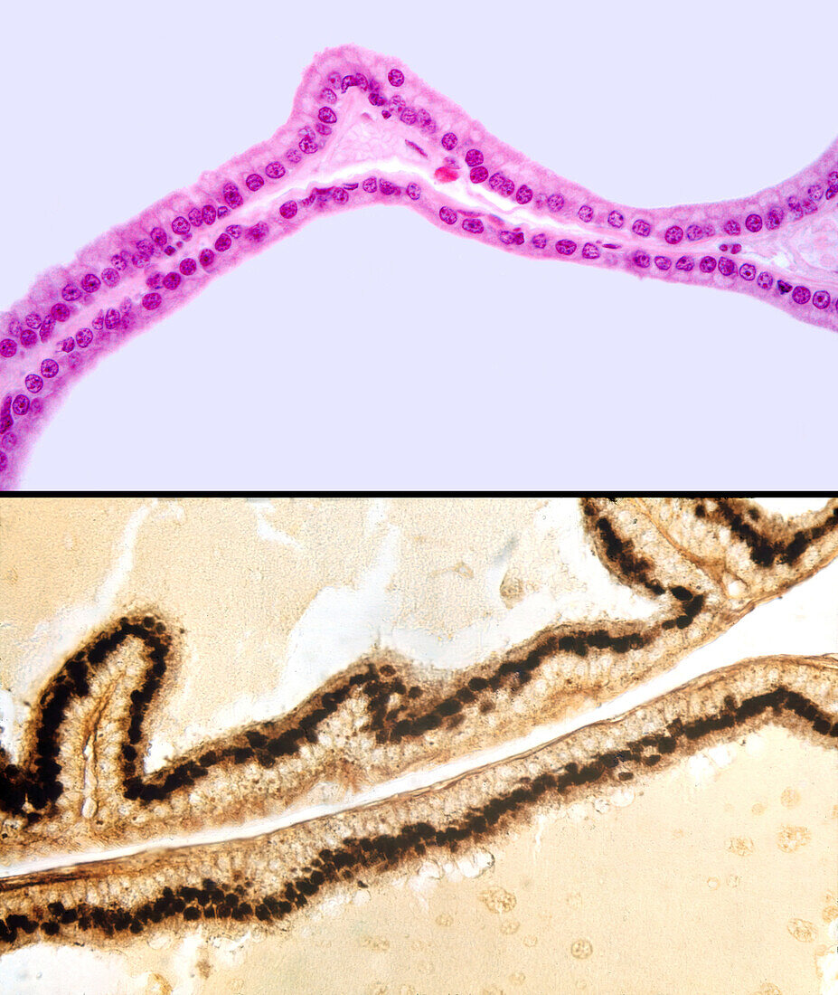
| Light micrograph of the septum between two alveoli of a rat coagulating gland lined by a simple columnar epithelium stained with haematoxylin and eosin (top) and the Da Fano silver method for Golgi apparatus (bottom). At top, epithelial cells show a supranuclear light band corresponding to the Golgi apparatus location (negative Golgi image). At bottom, these cells show a dark silver stained band above the nucleus corresponding to the location of the Golgi apparatus. | |
| Licence : | Droits gérés |
| Crédit: | Science Photo Library / JOSE CALVO |
| Taille de l’image : | 3840 px × 4556 px |
| Model Release : | Non requis |
| Property Release : | Non requis |
| Restrictions : | - |
Prix pour cette image À partir de 45 €
Produit vendu
(Calendrier, Carte postale, Carte de vœux, Impression sur textile, Packaging etc)
À partir de 45 €
Usage commercial
(Affichage, Annonce presse, Annonce TV, Carte, Digital - hors rés. sociaux, Digital - rés. sociaux etc)
À partir de 45 €
Éditorial
(Digital, Journal, Livre, Livre pratique, Magazine, Télévision etc)
À partir de 60 €
Usage non-commercial
(Digital - hors rés. sociaux, Digital - rés. sociaux etc)
À partir de 120 €
Mots clés
- appareil de Golgi,
- argent,
- aucun,
- en bonne santé,
- éosine,
- épithélium cylindrique,
- génital,
- Golgi,
- Golgi apparatus,
- hématoxyline,
- histologie,
- histologique,
- masculin,
- micrographie optique,
- microscope,
- microscope optique,
- microscopie,
- microscopie optique,
- normal,
- personne,
- sain,
- tissus,
- vésicule séminale