Fat cells, light micrograph
Numéro d’image : 13732516
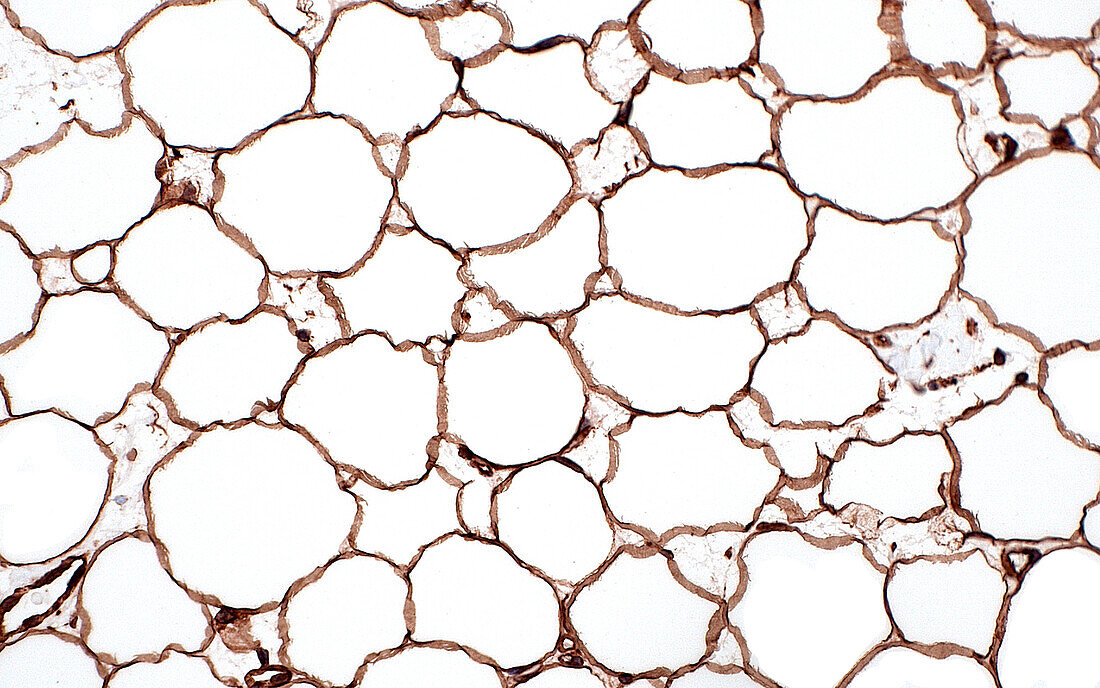
| Light micrograph of adipocytes (fat cells) stained by immunohistochemistry, a technique where antibodies detect specific proteins in tissue by attaching to them and giving off a certain colour. Here, the antibody used detected the protein vimentin, a protein commonly present in most cell types. The brown colour of the stain highlights the membranes of the fat cells. The fat cells themselves are mostly made up of the white spaces which is where lipids (fat) is normally stored. Haematoxylin and eosin stained tissue section. Magnification: 200x when printed at 10cm. | |
| Licence : | Droits gérés |
| Crédit: | Science Photo Library / ZIAD M. EL-ZAATARI |
| Taille de l’image : | 5000 px × 3125 px |
| Model Release : | Non requis |
| Property Release : | Non requis |
| Restrictions : | - |
Prix pour cette image À partir de 45 €
Produit vendu
(Calendrier, Carte postale, Carte de vœux, Impression sur textile, Packaging etc)
À partir de 45 €
Usage commercial
(Affichage, Annonce presse, Annonce TV, Carte, Digital - hors rés. sociaux, Digital - rés. sociaux etc)
À partir de 45 €
Éditorial
(Digital, Journal, Livre, Livre pratique, Magazine, Télévision etc)
À partir de 60 €
Usage non-commercial
(Digital - hors rés. sociaux, Digital - rés. sociaux etc)
À partir de 120 €
Mots clés
- adipocytes,
- agrandissement,
- anatomie,
- anatomique,
- arrière plan blanc,
- arrière-plan blanc,
- biologie,
- biologique,
- brun,
- cellules,
- corps humain,
- en bonne santé,
- fond blanc,
- glisser,
- graisse,
- histologie,
- histologique,
- métabolisme,
- micrographie optique,
- microscope,
- microscope optique,
- microscopie,
- microscopie optique,
- normal,
- sain,
- tissu mou,
- tissus