Seminal vesicle muscle wall, light micrograph
Numéro d’image : 13732483
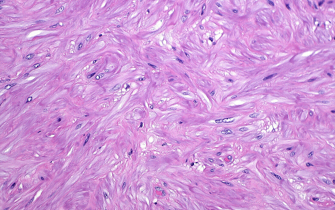
| Light micrograph of smooth muscle cells in the wall of a seminal vesicle (an organ next to the prostate gland). The pink parts are the cytoplasm of the cells and the dark blue ovoid structures are the cells’ nuclei. Haematoxylin and eosin stained tissue section. Magnification: 200x when printed at 10cm. | |
| Licence : | Droits gérés |
| Crédit: | Science Photo Library / ZIAD M. EL-ZAATARI |
| Taille de l’image : | 5000 px × 3127 px |
| Model Release : | Non requis |
| Property Release : | Non requis |
| Restrictions : | - |
Prix pour cette image À partir de 45 €
Produit vendu
(Calendrier, Carte postale, Carte de vœux, Impression sur textile, Packaging etc)
À partir de 45 €
Usage commercial
(Affichage, Annonce presse, Annonce TV, Carte, Digital - hors rés. sociaux, Digital - rés. sociaux etc)
À partir de 45 €
Éditorial
(Digital, Journal, Livre, Livre pratique, Magazine, Télévision etc)
À partir de 60 €
Usage non-commercial
(Digital - hors rés. sociaux, Digital - rés. sociaux etc)
À partir de 120 €
Mots clés
- agrandissement,
- anatomie,
- anatomique,
- biologie,
- biologique,
- cellules,
- coloration à l'hématoxyline et à l'éoisine,
- coloration HE,
- corps humain,
- en bonne santé,
- glisser,
- histologie,
- histologique,
- micrographie optique,
- microscope,
- microscope optique,
- microscopie,
- microscopie optique,
- muscle lisse,
- normal,
- rose,
- sain,
- système reproducteur masculin,
- tissus,
- vésicule séminale