Stomach glands, light micrograph
Numéro d’image : 13732456
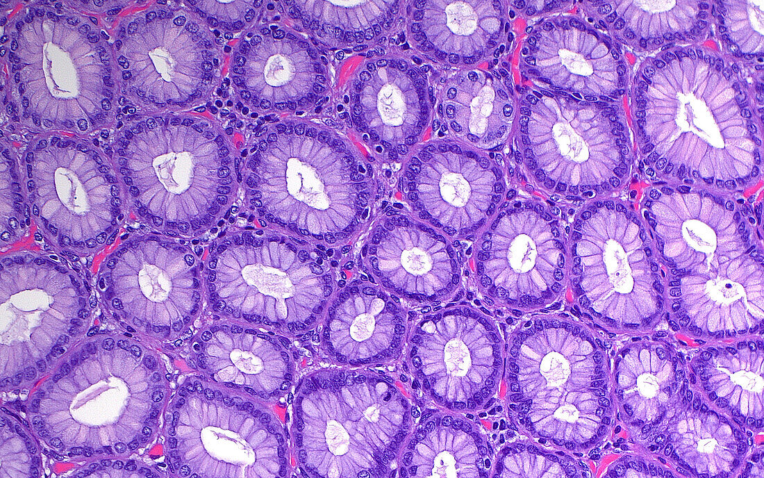
| Light micrograph of the lining cells of the stomach, seen in cross-section. Each of the circular structures represents a microscopic stomach gland, and there are capillaries (small blood vessels) squeezed in between each of the circular gland cross-sections. Haematoxylin and eosin stained tissue section. Magnification: 200x when printed at 10cm. | |
| Licence : | Droits gérés |
| Crédit: | Science Photo Library / ZIAD M. EL-ZAATARI |
| Taille de l’image : | 5000 px × 3125 px |
| Model Release : | Non requis |
| Property Release : | Non requis |
| Restrictions : | - |
Prix pour cette image À partir de 45 €
Produit vendu
(Calendrier, Carte postale, Carte de vœux, Impression sur textile, Packaging etc)
À partir de 45 €
Usage commercial
(Affichage, Annonce presse, Annonce TV, Carte, Digital - hors rés. sociaux, Digital - rés. sociaux etc)
À partir de 45 €
Éditorial
(Digital, Journal, Livre, Livre pratique, Magazine, Télévision etc)
À partir de 60 €
Usage non-commercial
(Digital - hors rés. sociaux, Digital - rés. sociaux etc)
À partir de 120 €
Mots clés
- agrandissement,
- anatomie,
- anatomique,
- biologie,
- biologique,
- cellules,
- coloration à l'hématoxyline et à l'éoisine,
- coloration HE,
- corps humain,
- digestion,
- en bonne santé,
- epithelium,
- épithélium,
- estomac,
- gastroentérologie,
- glisser,
- histologie,
- histologique,
- micrographie optique,
- microscope,
- microscope optique,
- microscopie,
- microscopie optique,
- normal,
- sain,
- système digestif,
- tissus