Bladder cancer cells with mitoses, light micrograph
Numéro d’image : 13732416
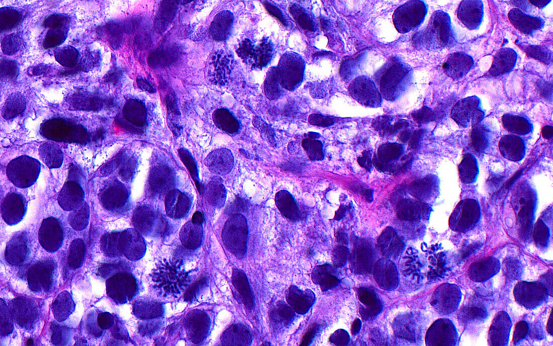
| Light micrograph of mitotic figures. A few mitotic figures are seen amongst the many cancerous cells in this image of bladder cancer. The mitoses are dividing cells and are identified by their many small linear structures which are the condensed chromosomes of the cells. Haematoxylin and eosin stained tissue section. Magnification: 400x when printed at 10 cm. | |
| Licence : | Droits gérés |
| Crédit: | Science Photo Library / ZIAD M. EL-ZAATARI |
| Taille de l’image : | 5000 px × 3128 px |
| Model Release : | Non requis |
| Property Release : | Non requis |
| Restrictions : | - |
Prix pour cette image À partir de 45 €
Produit vendu
(Calendrier, Carte postale, Carte de vœux, Impression sur textile, Packaging etc)
À partir de 45 €
Usage commercial
(Affichage, Annonce presse, Annonce TV, Carte, Digital - hors rés. sociaux, Digital - rés. sociaux etc)
À partir de 45 €
Éditorial
(Digital, Journal, Livre, Livre pratique, Magazine, Télévision etc)
À partir de 60 €
Usage non-commercial
(Digital - hors rés. sociaux, Digital - rés. sociaux etc)
À partir de 120 €
Mots clés
- agrandissement,
- anatomie,
- anormal,
- cancer de la vessie,
- cellules,
- coloration à l'hématoxyline et à l'éoisine,
- coloration HE,
- corps humain,
- croissance de tumeur,
- désordre,
- diagnostic,
- diagnostique,
- état,
- glisser,
- histologie,
- histologique,
- histopathologie,
- maladie,
- médecine,
- micrographie optique,
- microscope,
- microscope optique,
- microscopie,
- microscopie optique,
- oncologie,
- pathologie,
- tissus,
- trouble,
- vessie