Lymphocytic colitis, light micrograph
Numéro d’image : 13732412
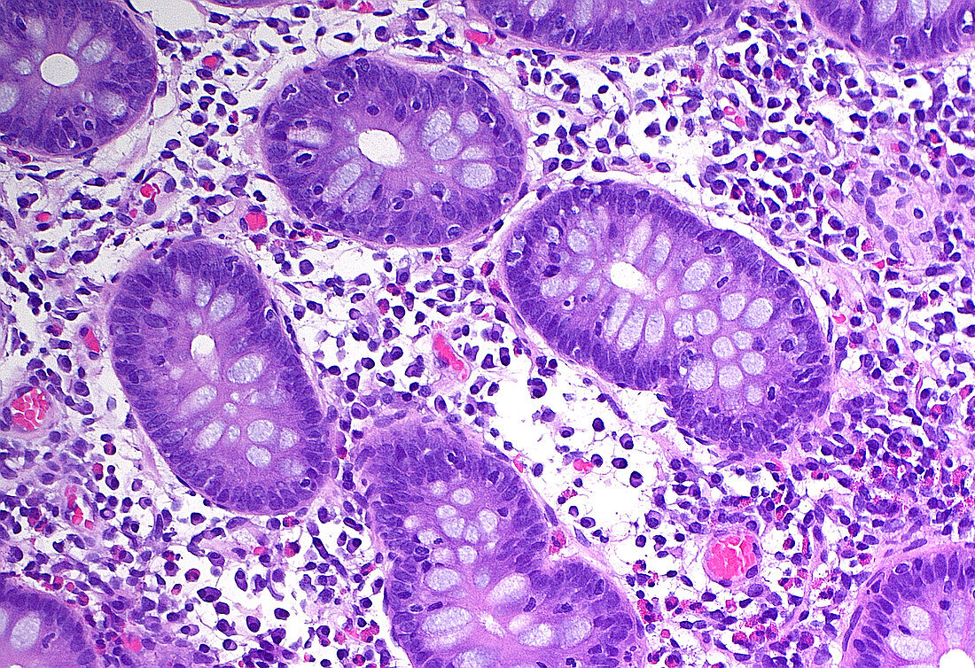
| Light micrograph of lymphocytic colitis, which is diagnosed by a pathologist on the basis of increased lymphocytes within colon epithelium. The colon crypts are the roughly oval or tubular structures, four of which are around the centre of this image. Note the many small blue dots, which are the lymphocytes within the crypts. Haematoxylin and eosin stained tissue section. Magnification: 200x when printed at 10 cm. | |
| Licence : | Droits gérés |
| Crédit: | Science Photo Library / ZIAD M. EL-ZAATARI |
| Taille de l’image : | 5000 px × 3427 px |
| Model Release : | Non requis |
| Property Release : | Non requis |
| Restrictions : | - |
Prix pour cette image À partir de 45 €
Produit vendu
(Calendrier, Carte postale, Carte de vœux, Impression sur textile, Packaging etc)
À partir de 45 €
Usage commercial
(Affichage, Annonce presse, Annonce TV, Carte, Digital - hors rés. sociaux, Digital - rés. sociaux etc)
À partir de 45 €
Éditorial
(Digital, Journal, Livre, Livre pratique, Magazine, Télévision etc)
À partir de 60 €
Usage non-commercial
(Digital - hors rés. sociaux, Digital - rés. sociaux etc)
À partir de 120 €
Mots clés
- agrandissement,
- anatomie,
- anormal,
- cellules,
- colon,
- coloration à l'hématoxyline et à l'éoisine,
- coloration HE,
- corps humain,
- désordre,
- diagnostic,
- diagnostique,
- endoscopie,
- état,
- gastroentérologie,
- glisser,
- histologie,
- histologique,
- histopathologie,
- inflammation,
- maladie,
- médecine,
- micrographie optique,
- microscope,
- microscope optique,
- microscopie,
- microscopie optique,
- pathologie,
- tissus,
- trouble