Heart muscle cells, light micrograph
Numéro d’image : 13732397
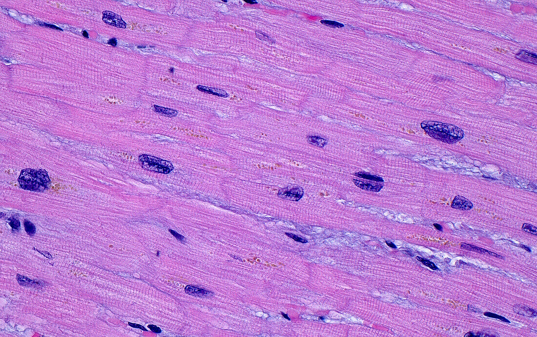
| Light micrograph of cardiomyocytes (heart muscle cells). The heart muscle cells show many fine parallel fibres and intercalated discs (short squiggly light pink lines). The nuclei (dark blue circular structures) are slightly enlarged or hypertrophic, a result of chronic stress or damage on the heart. There is also fine yellowish lipofuscin pigment (small yellow-orange dots), a normal finding especially in older people. Haematoxylin and eosin stained tissue section. Magnification: 400x when printed at 10 cm. | |
| Licence : | Droits gérés |
| Crédit: | Science Photo Library / ZIAD M. EL-ZAATARI |
| Taille de l’image : | 5000 px × 3138 px |
| Model Release : | Non requis |
| Property Release : | Non requis |
| Restrictions : | - |
Prix pour cette image À partir de 45 €
Produit vendu
(Calendrier, Carte postale, Carte de vœux, Impression sur textile, Packaging etc)
À partir de 45 €
Usage commercial
(Affichage, Annonce presse, Annonce TV, Carte, Digital - hors rés. sociaux, Digital - rés. sociaux etc)
À partir de 45 €
Éditorial
(Digital, Journal, Livre, Livre pratique, Magazine, Télévision etc)
À partir de 60 €
Usage non-commercial
(Digital - hors rés. sociaux, Digital - rés. sociaux etc)
À partir de 120 €
Mots clés
- agrandissement,
- anatomie,
- cardiomyocytes,
- cardiovasculaire,
- cellules,
- cœur,
- coloration à l'hématoxyline et à l'éoisine,
- coloration HE,
- corps humain,
- désordre,
- diagnostic,
- diagnostique,
- disques intercalatés,
- état,
- glisser,
- histologie,
- histologique,
- histopathologie,
- maladie,
- médecine,
- micrographie optique,
- microscope,
- microscope optique,
- microscopie,
- microscopie optique,
- muscle,
- pathologie,
- tissus,
- trouble