Fat necrosis, light micrograph
Numéro d’image : 13732389
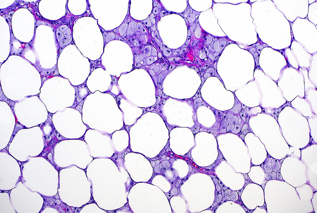
| Light micrograph of fat necrosis. Dying fat cells (the white circular spaces) have no nuclei and are admixed with foamy histiocytes (the blue-gray cells in between). Haematoxylin and eosin stained tissue section. Magnification: 100x when printed at 10 cm. | |
| Licence : | Droits gérés |
| Crédit: | Science Photo Library / ZIAD M. EL-ZAATARI |
| Taille de l’image : | 5000 px × 3373 px |
| Model Release : | Non requis |
| Property Release : | Non requis |
| Restrictions : | - |
Prix pour cette image À partir de 45 €
Produit vendu
(Calendrier, Carte postale, Carte de vœux, Impression sur textile, Packaging etc)
À partir de 45 €
Usage commercial
(Affichage, Annonce presse, Annonce TV, Carte, Digital - hors rés. sociaux, Digital - rés. sociaux etc)
À partir de 45 €
Éditorial
(Digital, Journal, Livre, Livre pratique, Magazine, Télévision etc)
À partir de 60 €
Usage non-commercial
(Digital - hors rés. sociaux, Digital - rés. sociaux etc)
À partir de 120 €
Mots clés
- agrandissement,
- anatomie,
- anormal,
- cellules,
- coloration à l'hématoxyline et à l'éoisine,
- coloration HE,
- corps humain,
- désordre,
- diagnostic,
- diagnostique,
- état,
- glisser,
- graisse,
- histiocytes,
- histologie,
- histologique,
- histopathologie,
- inflammation,
- maladie,
- médecine,
- micrographie optique,
- microscope,
- microscope optique,
- microscopie,
- microscopie optique,
- pathologie,
- tissu mou,
- tissus,
- trouble