Venule, light micrograph
Numéro d’image : 13686897
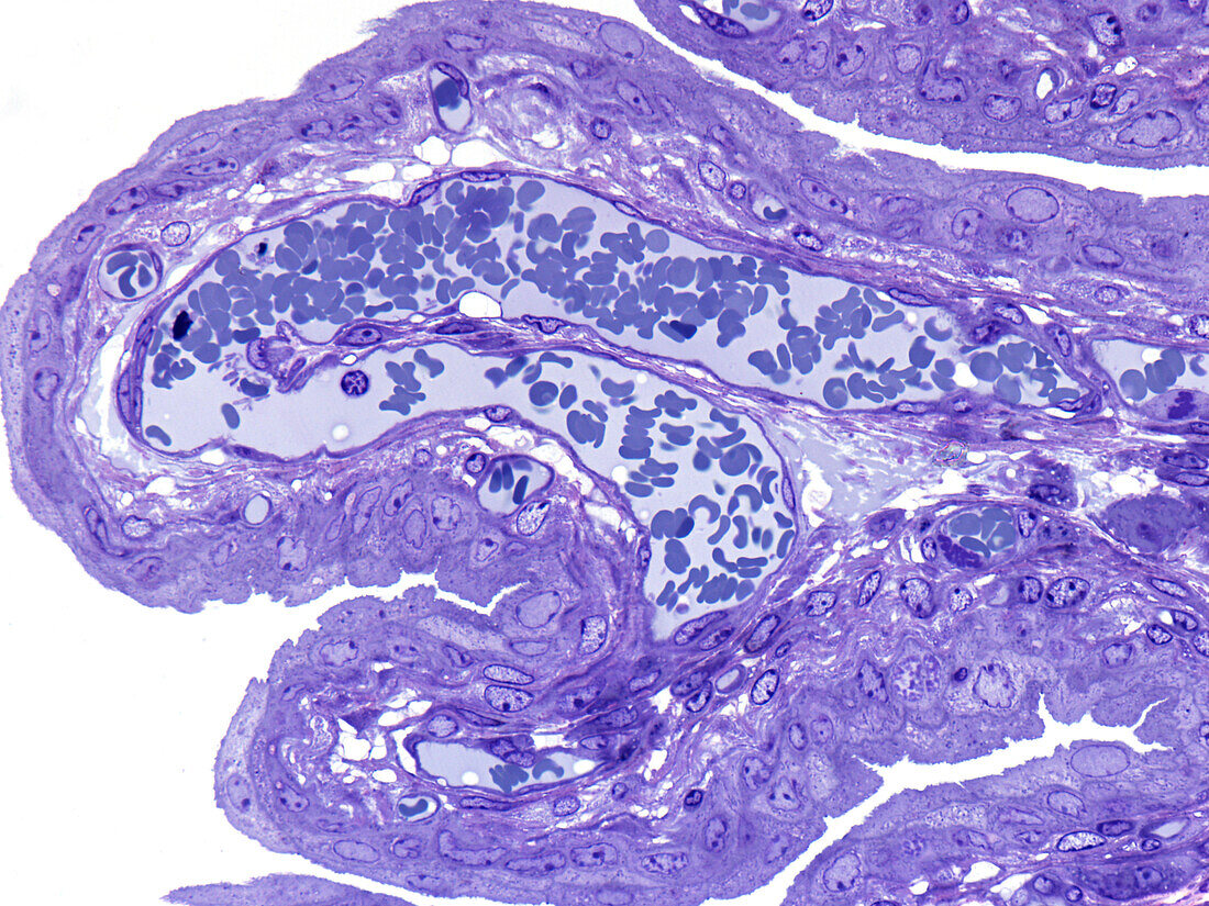
| Venule, light micrograph. A venule in a U-shaped profile is shown adjacent to the surface epithelium of the urinary bladder. The venule shows a very thin wall comprised of endothelium. A number of capillaries containing erythrocytes (blue) are noted in the connective tissue. Epoxy resin section, Toluidine blue stain. Magnification: x350 when width printed at 10cm. | |
| Licence : | Droits gérés |
| Crédit: | Science Photo Library / Microscape |
| Taille de l’image : | 4843 px × 3632 px |
| Model Release : | Non requis |
| Property Release : | Non requis |
| Restrictions : | - |
Prix pour cette image À partir de 45 €
Produit vendu
(Calendrier, Carte postale, Carte de vœux, Impression sur textile, Packaging etc)
À partir de 45 €
Usage commercial
(Affichage, Annonce presse, Annonce TV, Carte, Digital - hors rés. sociaux, Digital - rés. sociaux etc)
À partir de 45 €
Éditorial
(Digital, Journal, Livre, Livre pratique, Magazine, Télévision etc)
À partir de 60 €
Usage non-commercial
(Digital - hors rés. sociaux, Digital - rés. sociaux etc)
À partir de 120 €