Human ureter, light micrograph
Numéro d’image : 13673515
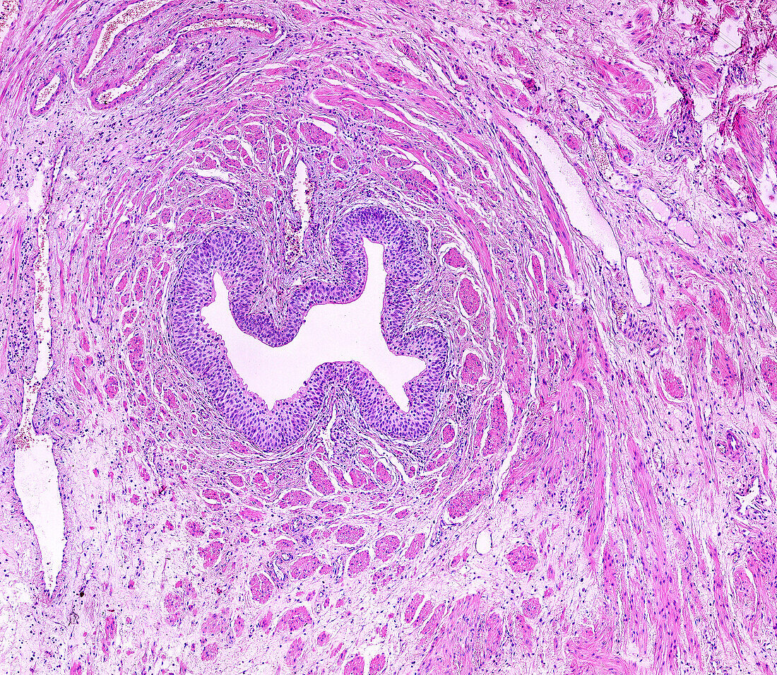
| Light micrograph of a cross-sectioned human ureter stained with haematoxylin and eosin. Lining the lumen is the folded mucosa, formed by a transitional epithelium, or urothelium, with several layers of cells and the connective tissue lamina propria. Surrounding this are the muscular layers formed by fascicles of smooth muscle fibres that, unlike in the digestive tube, are not clearly arranged in layers. | |
| Licence : | Droits gérés |
| Crédit: | Science Photo Library / JOSE CALVO |
| Taille de l’image : | 7719 px × 6698 px |
| Model Release : | Non requis |
| Property Release : | Non requis |
| Restrictions : | - |
Prix pour cette image À partir de 45 €
Produit vendu
(Calendrier, Carte postale, Carte de vœux, Impression sur textile, Packaging etc)
À partir de 45 €
Usage commercial
(Affichage, Annonce presse, Annonce TV, Carte, Digital - hors rés. sociaux, Digital - rés. sociaux etc)
À partir de 45 €
Éditorial
(Digital, Journal, Livre, Livre pratique, Magazine, Télévision etc)
À partir de 60 €
Usage non-commercial
(Digital - hors rés. sociaux, Digital - rés. sociaux etc)
À partir de 120 €
Mots clés
- arrière plan blanc,
- arrière-plan blanc,
- aucun,
- epithelium,
- épithélium,
- épithélium de transition,
- fond blanc,
- histologie,
- micrographie,
- micrographie optique,
- microscope,
- microscope optique,
- microscopie,
- microscopie optique,
- personne,
- rénale,
- système urinaire,
- transitoire,
- uretère,
- urologie,
- urothelium