Cross-section of the neck, light micrograph
Numéro d’image : 13673509
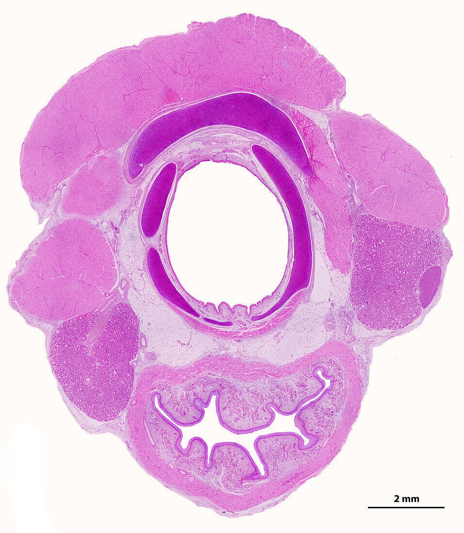
| Light micrograph showing a cross-section of the neck. At the top and sides are the striated muscles of the neck. Beneath the muscle, the largest cartilage is the thyroid cartilage. At centre is the trachea, showing C-shaped tracheal rings of hyaline cartilage. The tracheal mucosa is located inside the cartilage. On the sides, behind the muscles are two thyroid lobules. The right thyroid lobule shows a small parathyroid gland. At bottom there is a cross-sectioned oesophagus with a folded lumen. The spaces among these structures are filled with adipose (fat) tissue. The scale bar corresponds to 2 mm. | |
| Licence : | Droits gérés |
| Crédit: | Science Photo Library / JOSE CALVO |
| Taille de l’image : | 3342 px × 3840 px |
| Model Release : | Non requis |
| Property Release : | Non requis |
| Restrictions : | - |
Prix pour cette image À partir de 45 €
Produit vendu
(Calendrier, Carte postale, Carte de vœux, Impression sur textile, Packaging etc)
À partir de 45 €
Usage commercial
(Affichage, Annonce presse, Annonce TV, Carte, Digital - hors rés. sociaux, Digital - rés. sociaux etc)
À partir de 45 €
Éditorial
(Digital, Journal, Livre, Livre pratique, Magazine, Télévision etc)
À partir de 60 €
Usage non-commercial
(Digital - hors rés. sociaux, Digital - rés. sociaux etc)
À partir de 120 €
Mots clés
- adipeux,
- anneaux,
- arrière plan blanc,
- arrière-plan blanc,
- aucun,
- cartilage,
- cartilage thyroïdien,
- fond blanc,
- histologie,
- histologique,
- hyalin,
- micrographie,
- micrographie optique,
- microscope,
- microscope optique,
- microscopie,
- microscopie optique,
- microscopique,
- mucosa,
- muqueuse,
- œsophage,
- oesophagus,
- parathyroïde,
- personne,
- presque transparent,
- respiratoire,
- thyroïde,
- trachea,
- trachée,
- translucide