Thymus, light micrograph
Numéro d’image : 13673500
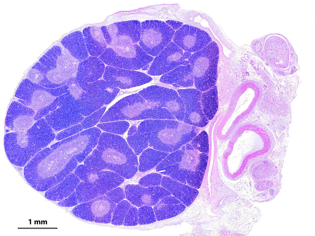
| Light micrograph showing a young thymus. The organization into lobules is clearly seen. In each lobule, the peripheral cortex appears more stained, due to the high density of T-lymphocyte precursor cells. In the paler centre of each lobule there are many Hassall's corpuscles. On the right side of the thymus there are blood vessels and nerves. The scale bar corresponds to 1 mm. | |
| Licence : | Droits gérés |
| Crédit: | Science Photo Library / JOSE CALVO |
| Taille de l’image : | 4099 px × 3072 px |
| Model Release : | Non requis |
| Property Release : | Non requis |
| Restrictions : | - |
Prix pour cette image À partir de 45 €
Produit vendu
(Calendrier, Carte postale, Carte de vœux, Impression sur textile, Packaging etc)
À partir de 45 €
Usage commercial
(Affichage, Annonce presse, Annonce TV, Carte, Digital - hors rés. sociaux, Digital - rés. sociaux etc)
À partir de 45 €
Éditorial
(Digital, Journal, Livre, Livre pratique, Magazine, Télévision etc)
À partir de 60 €
Usage non-commercial
(Digital - hors rés. sociaux, Digital - rés. sociaux etc)
À partir de 120 €
Mots clés
- arrière plan blanc,
- arrière-plan blanc,
- aucun,
- cortex,
- fond blanc,
- globules,
- histologie,
- histologique,
- humain,
- immunitaire,
- immunologie,
- immunologique,
- lymphocyte,
- medulla,
- médullaire,
- micrographie,
- micrographie optique,
- microscope,
- microscope optique,
- microscopie,
- microscopie optique,
- personne,
- système immunitaire,
- thymus