Human pineal gland, light micrograph
Numéro d’image : 13673487
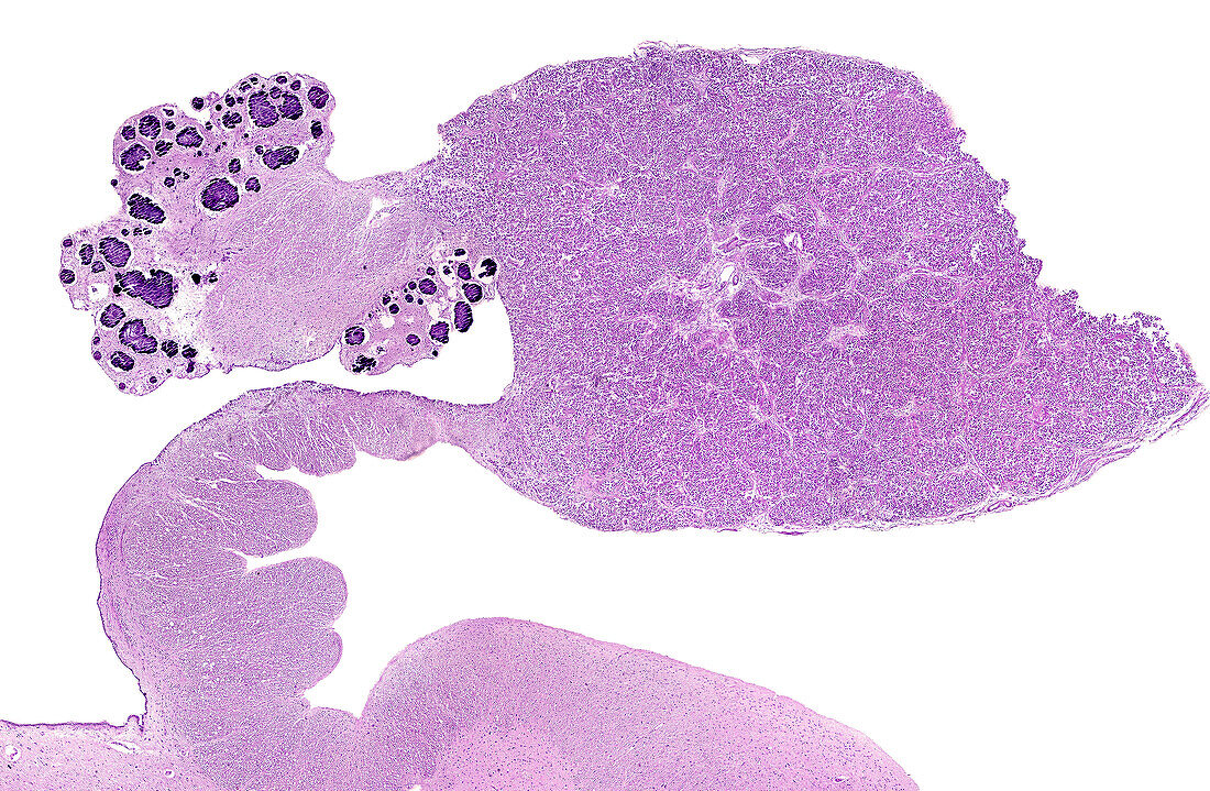
| Light micrograph showing a sagittal section of a human pineal gland. The sample is from a 69-year-old man. The pineal recess is on the left, opened to the third ventricle and situated between the two commissures. The habenular commissure shows a huge deposit of calcium concretions (corpora arenacea, brain sand). On the contrary, the posterior commissure is free of them. The pineal parenchyma shows a distinct lobulation and some small glial spots. | |
| Licence : | Droits gérés |
| Crédit: | Science Photo Library / JOSE CALVO |
| Taille de l’image : | 3840 px × 2510 px |
| Model Release : | Non requis |
| Property Release : | Non requis |
| Restrictions : | - |
Prix pour cette image À partir de 45 €
Produit vendu
(Calendrier, Carte postale, Carte de vœux, Impression sur textile, Packaging etc)
À partir de 45 €
Usage commercial
(Affichage, Annonce presse, Annonce TV, Carte, Digital - hors rés. sociaux, Digital - rés. sociaux etc)
À partir de 45 €
Éditorial
(Digital, Journal, Livre, Livre pratique, Magazine, Télévision etc)
À partir de 60 €
Usage non-commercial
(Digital - hors rés. sociaux, Digital - rés. sociaux etc)
À partir de 120 €
Mots clés
- anatomie,
- arrière plan blanc,
- arrière-plan blanc,
- aucun,
- corps humain,
- endocrine,
- épiphyse,
- fond blanc,
- glande endocrinale,
- glande endocrine,
- glande pinéale,
- histologie,
- histologique,
- hormonal,
- hormonale,
- hormone,
- humain,
- mélatonine,
- micrographie,
- micrographie optique,
- microscope,
- microscope optique,
- microscopie,
- microscopie optique,
- personne,
- pinéal,
- rythme circadien