Developing long bone, light micrograph
Numéro d’image : 13673460
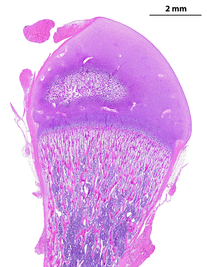
| Light micrograph showing a developing long bone (femur, or thigh bone). At top, the epiphysis is made by hyaline cartilage that shows an initial secondary ossification centre. At bottom is the secondary diaphyseal ossification centre. The round ligament can be seen at top left. The scale bar corresponds to 2 mm. | |
| Licence : | Droits gérés |
| Crédit: | Science Photo Library / JOSE CALVO |
| Taille de l’image : | 2978 px × 3840 px |
| Model Release : | Non requis |
| Property Release : | Non requis |
| Restrictions : | - |
Prix pour cette image À partir de 45 €
Produit vendu
(Calendrier, Carte postale, Carte de vœux, Impression sur textile, Packaging etc)
À partir de 45 €
Usage commercial
(Affichage, Annonce presse, Annonce TV, Carte, Digital - hors rés. sociaux, Digital - rés. sociaux etc)
À partir de 45 €
Éditorial
(Digital, Journal, Livre, Livre pratique, Magazine, Télévision etc)
À partir de 60 €
Usage non-commercial
(Digital - hors rés. sociaux, Digital - rés. sociaux etc)
À partir de 120 €